Interventional Pulmonology for Lung Cancer
How is Interventional Pulmonology Different at Hoag?
Hoag has been Orange County’s leader in the advanced diagnosis and treatment of lung cancer for decades. With the introduction of our new Interventional Pulmonology program, we take that commitment to excellence to the next level. At Hoag, our team includes Javier A. Longoria, M.D., the area’s only practicing Interventional Pulmonologist, and the next-generation treatment and interventional options that can make a real difference in outcomes, recoveries and patients’ lives. Read more about Dr. Longoria at the bottom of this page.
FULLY-INTEGRATED TEAMS AND TREATMENT
One important way Interventional Pulmonology at Hoag is different is that our IP team is fully integrated with the Hoag Family Cancer Institute. That integration gives our Interventional Pulmonology program instant access and communication with the area’s most experienced and highly-trained thoracic surgeons, radiation oncologists and other specialists. Collectively, they’ve spent decades studying and treating lung cancer, including contributing to many of the treatments and minimally-invasive techniques used around the world. The result: a team that’s always looking for the best and most appropriate path forward for every patient, from diagnosis to recovery.
What is Interventional Pulmonology?
Pulmonology is a field of medicine that specializes in the diagnosis and treatment of lung cancer and other airway disorders related to breathing and the respiratory system, including the lungs, airways, breathing-related blood vessels and thoracic cavity, which is the area inside your upper chest that contains the lungs, heart and other organs. In the past, treating these conditions often required invasive surgery that could extend recovery time to weeks or months.
Interventional Pulmonology is a different approach to treating and managing lung cancer and other conditions of the respiratory system. While still a relatively new field, Interventional Pulmonology is dedicated to less-invasive methods of diagnosing, treating and managing lung cancer and its symptoms.
Rather than major surgery, Interventional Pulmonology utilizes next-generation imaging and techniques like advanced, guided bronchoscopy to improve outcomes and shorten hospital stays and recovery times. These efforts are overseen by a team of oncologists, thoracic surgeons and other specialists who are highly-trained in this less-invasive approach.
At Hoag, advanced techniques utilized by our Interventional Pulmonology program include advanced endoscopic surgery, which is performed by passing small instruments and cameras into the thoracic cavity through small incisions, and robotic-assisted bronchoscopy with three-dimensional guidance, in which a camera is passed down the throat and into the respiratory system so the lungs and airways can be directly viewed by physicians.
Lung Cancer Treatment through Interventional Pulmonology?
Interventional Pulmonology at Hoag is an important part of the multidisciplinary approach to diagnosing and treating the complexities of lung cancer. By utilizing minimally invasive techniques, interventional pulmonologists can provide accurate diagnoses and effective treatments while minimizing discomfort and improving outcomes for our patients.
TOMORROW’S TECHNIQUES, TODAY
Interventional Pulmonology is about responsibly pushing the boundaries of medicine and techniques to help patients in ways that would have been simply impossible even a few years ago. Among the next-generation approaches employed by Interventional Pulmonology at Hoag:
- Cios Spin: a new imaging technology for diagnosing lung cancer earlier, more accurately and less invasively. Hoag is the first hospital in Orange County to offer this technology.
- The Ion by Intuitive Robotic-Assisted Bronchoscopy Platform: Hoag is proud to be one of the few Interventional Pulmonology programs in the U.S. to offer Ion Robotic-Assisted Bronchoscopy. It’s a robot-enabled technique that allows for safe, minimally-invasive collection of tissue samples for biopsy by passing instruments down the windpipe, even when lesions are deep in the lungs. Ion’s thin and maneuverable catheter allows clinicians to reach small lesions in all 18 segments of the lung, without invasive surgery or needle biopsies that puncture the skin.
- Cone-Beam Computed Tomography (CBCT) with 3D Fluoroscopy: Hoag is one of the few centers in the nation to offer Cone-Beam Computed Tomography (CBCT) with 3D Fluoroscopy for lung conditions. It’s an advanced imaging technique that creates three-dimensional (3D) images of tissues inside the body. This enhanced imaging allows for easier treatment and ablation of tumors of the lung. In addition to utilizing radiation exposures that are up to 10 times less than conventional CT scans, CBCT is also much faster than conventional CT.
- Convex-Probe Endobronchial Ultrasound Bronchoscopy (CP-EBUS), which is a revolutionary form of bronchoscopy that incorporates a convex ultrasound transducer into the tip of the bronchoscope, which is a small, flexible camera passed down the throat and into the airways. Incorporating ultrasound gives specialists real-time guidance when obtaining tissue samples from deep in the lungs, allowing sampling to be more precise. Though CP-EBUS allows advanced diagnosis of different types of pulmonary disorders, including infections and inflammation, it is particularly useful in the diagnosis and staging of lung cancer.
- Radial-Probe Endobronchial Ultrasound Bronchoscopy (RP-EBUS), which is another advanced, highly-accurate form of guided bronchoscopy. Unlike CP-EBUS, RP-EBUS involves a rotating ultrasound source that’s passed through the bronchoscope. This allows pulmonary nodules and other issues to be detected with much greater accuracy, without the need for radiation-based imaging.
- Cryoablation, which is a technique that utilizes localized, extreme cold to treat certain cancers, including lung cancer that may have been deemed inoperable. During the procedure, a thin instrument known as a cryoprobe is inserted through a bronchoscope until it comes in direct contact with the cancerous or benign tumor or lesion. Gas is then used to reduce the tip of the cryoprobe to below freezing, destroying cancerous cells through repeated freezing and thawing cycles.
If you have been diagnosed or are concerned that you may have Lung Cancer, trust Hoag to be with you every step of the way offering the expertise of a multidisciplinary team and the latest diagnostic and treatment options to care for you. At Hoag, you are not alone.
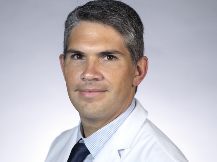
Javier A. Longoria, M.D.
Dr. Longoria attended medical school at Loyola University Chicago, Stritch School of Medicine. He completed his residency in internal medicine and fellowship training in both interventional pulmonology and pulmonary and critical care medicine at University of California, Irvine. He is board certified in internal medicine, pulmonary medicine and critical care medicine.
Learn More