
Hoag Memorial Hospital Presbyterian
1 Hoag Drive, Building #41
Newport Beach, CA 92663
949-722-6237
Hoag Radiation Oncology Program is a technology-dependent medical specialty that requires sophisticated computers and software, and high-energy linear accelerators to deliver tailored therapeutic doses of radiation. Treatment complications and outcomes can be significantly improved by use of the right instrumentation and technological support. Our Radiation Oncologists and Medical Physicists use state-of-the-art resources to develop three-dimensional treatment plans that deliver precise radiation doses for each patient’s individual course of radiation therapy. Learn about many of our state-of-the-art technologies below.
The ViewRay MRIdian™ linear accelerator is the most advanced radiation oncology tool available. Through the generosity of community donors Dean and Gerda Koontz, Hoag was the second in California to acquire the MRIdian, and 16th in the nation. There has been a recent upgrade to this state-of-the-art system, and the MRidian now operates with an A3i software and hardware upgrade.
Unlike conventional linear accelerators, the ViewRay MRIdian utilizes MRI imaging in combination with a linear accelerator, which allows Hoag clinicians to obtain real-time, high resolution images of a patient’s tumor during treatment. If the tumor has shifted from movement in the bowels, or by a patient’s breath, radiation delivery stops to avoid radiating healthy tissue. This level of precision allows Hoag clinicians to deliver a higher, potentially more effective, radiation dose while sparing healthy surrounding tissue and improving upon side effects.
Traditionally, CT scans are taken before radiation treatment to help clinicians map out where to align the radiation beam during sessions. This type of imaging, however, does not usually take into account the normal functions of the body such as breathing or movement in the gastrointestinal tract, which can cause the tumor and/or normal tissues and organs to move from day to day during treatment. As a result, the healthy surrounding tissue are at a high risk of being damaged in the process.
MRI imaging allows Hoag clinicians to deliver targeted radiation, usually in fewer sessions, while sparing healthy surrounding tissue.
The ViewRay MRIdian also allows Hoag clinicians to more precisely and effectively treat tumors of the abdomen, like pancreas, liver and prostate. The MRI imaging provides high quality soft tissue definition, providing a clear treatment area.
Tomotherapy® is a specialized treatment machine specifically designed for Image-Guided Intensity-Modulated Radiation Therapy (IG-IMRT). The concept of treating cancer with radiation is a simple one: irradiate the cancer cells and they die. The challenge, therefore, lies in sparing the healthy tissue surrounding the tumor – that’s precisely what Tomotherapy is all about.
Tomotherapy achieves the ability to deliver maximum radiation dose to a target volume while sparing the surrounding healthy tissue by utilizing a CT scanner for daily image guidance to ensure proper patient set-up based on the internal anatomy. The machine’s superior ability to modulate the intensity of the radiation to conform around the target volume also significantly decreases the dose to healthy tissue.
“The breakthrough technology that makes Tomotherapy unique is that it combines the capabilities of a standard CT scanner with those of a radiation therapy linear accelerator,” explains Radiation Oncologist Peter Chen, M.D. “Like a standard CT scanner, the Tomotherapy source spirals around the patient like a corkscrew, enabling it to take three-dimensional pictures that help doctors to precisely localize treatments. However, instead of the imaging x-rays from a standard CT scanner, the Tomotherapy source, like a linear accelerator, emits cancer-killing megavoltage photons. With both these capabilities in hand, doctors are able to treat patients with unprecedented accuracy.”
Traditionally, radiation therapy treated a tumor by focusing relatively large beams of radiation from two to six directions. In contrast, Tomotherapy uses hundreds of pencil beams of radiation, rotating in a spiral around the tumor and hitting it from all directions. As the beam is rotating, the intensity of the radiation is varied, allowing the radiation oncologist to deliver the radiation with incredible precision. In addition, the radiation can be sculpted to fit the shape of the patient’s tumor, again providing more precise and effective treatment.
Another radiation treatment challenge lies in the movement of tumors – sometimes as little as a millimeter. The Tomotherapy system can provide 3D CT imaging immediately prior to treatment to verify the location of the patient’s tumor. By confirming the precise tumor location, the accuracy of the delivery is greatly increased.
Tomotherapy uses 3D Conformal Radiation Therapy (3DCRT) and IMRT technologies, with the addition of helical delivery and 3D imaging (IG-IMRT) for treatment verification.
Learn how Tomotherapy is used in breast cancer treatment.
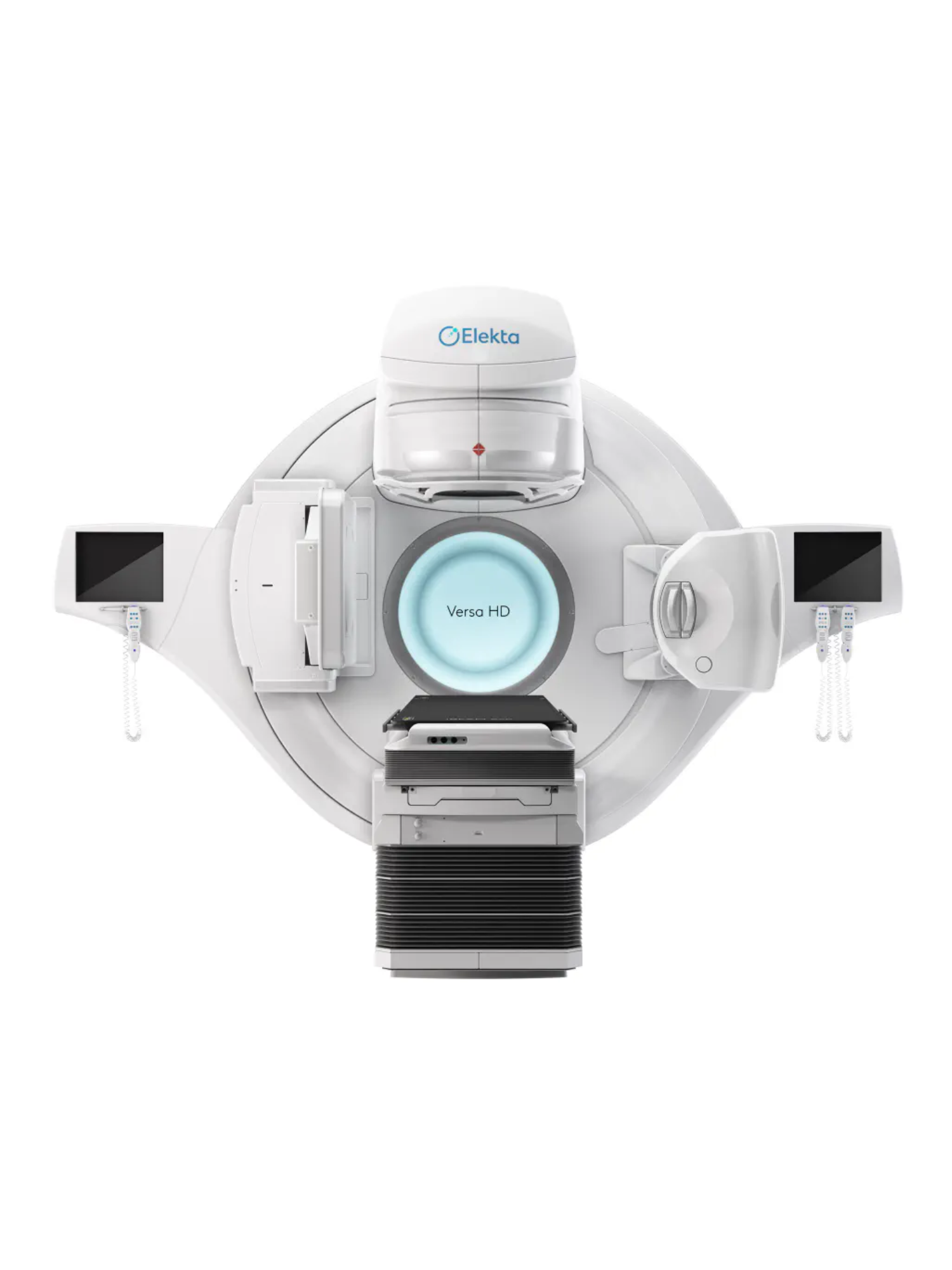
Hoag’s Elekta Versa HD is a state-of-the-art computer controlled linear accelerator with VMAT capabilities and the latest in 3D and 4D image guidance for increased accuracy. Equipped with both conebeam CT and megavoltage imaging the Versa HD can insure precise daily patient positioning and tumor targeting. Featuring an advanced MLC (multileaf collimator), Align RT surface guided technology, and a 6D Hexapod Evo RT system, the Elekta Versa HD represents one of latest advances in treatment technology.
Stereotactic Radiation Therapy (SRT) is an intermediate technique, with many of the characteristics of both Stereotactic Radiosurgery (SRS) and Image-Guided Intensity-Modulated Radiation Therapy (IG-IMRT). When applied outside of the brain, this technique is often called Stereotactic Body Radiation Therapy (SBRT). SRT uses doses higher than standard doses with conventional radiation therapy, but lower that SRS. The length of treatment is also intermediate – typically given in five daily dose fractions.
At Hoag, SRT and SBRT are delivered with the Viewray MRIdian, the Elekta Versa HD and the Tomotherapy unit. Each modality is especially suited for SRT/SBRT because of the precise nature of the imaging types and their ability to accurately identify and adapt to tumor changes. When delivered to the brain, SRT uses a thermoplastic immobilization mask that is molded to the patient’s head. In contrast to the rigid head frame of SRS, this mask is applied non-invasively. When delivered to the body (SBRT), the patient is placed in a molded cradle, and wrapped with plastic covering. Suction is applied to hold the patient in a stable position. In both circumstances, 3D imaging is performed prior to each treatment to verify correct positioning. SRT is used to treat benign and malignant tumors of the brain, such as meningiomas, pituitary adenomas, acoustic neuromas, large metastases (spread of cancer from other sites in the body), optic tumors, and gliomas (anaplasticastrocytoma, glioblastomamultiforme). SBRT is used to treat selected tumors in the body, such as spinal lesions, liver metastases and lung metastases.
Hoag has a distinct advantage with active SRT/SBRT and SRS programs in the same facility. Our weekly multidisciplinary Neuro-Oncology Tumor Board specialists are therefore able to recommend and carry out optimal treatment regimens within this fully integrated facility assuring the patients the best possible treatment options.
Intensity-Modulated Radiation Therapy (IMRT) uses intricately shaped beams to enhance the techniques used in 3D Conformal Radiation Therapy (3DCRT). In 3DCRT, intensity of the radiation is uniform throughout the shape of the beam. With IMRT, the radiation intensity is non-uniform throughout the beam shape. This non-uniform intensity allows for better control in shaping the radiation delivered to the target volume, while avoiding healthy tissue. (For example, when treating a head and neck cancer while trying to avoid nearby salivary gland and spinal cord.) The complex shaping of radiation using IMRT adds to the capabilities of 3DCRT, and allows the radiation oncologist to give more dose to the tumor while limiting exposure of normal tissue to safe levels.
IMRT can be further enhanced with the use of image-guidance (IG-IMRT). One problem that a radiation oncologist faces is how to position the patients properly for their daily treatments. Tumors aren’t always where they are expected because of movement with breathing, expansion of the gastrointestinal tract with air or other similar factors. In IG-IMRT, an image is taken daily prior to the radiation treatment, and changes in set up are made to ensure that the tumor is targeted appropriately. (For example, to help visualize the target, the radiation oncologist may implant metal markers in the prostate gland.) Our Varian 21EX uses a digital imager to visualize metal markers in a 2D fashion on a daily basis. Alternatively, some machines, such as Tomotherapy, can take a 3D image that shows the tumor and the surrounding anatomy directly in order to achieve the appropriate set up. Such image-guided techniques make treatment more accurate, ensuring that radiation is directed at the appropriate target and away from the adjacent healthy tissue.
High Dose Rate Brachytherapy (HDR), also referred to as “internal radiation therapy” is a radiation treatment allowing a small radioactive source to be temporarily placed inside numerous types of tumors.
Under computer control the position and timing of source placement can be precisely controlled, allowing the physician to shape the radiation dose to the target. Because of the high dose rate characteristics, brachytherapy treatments can often be delivered on an outpatient basis or with a minimal hospital stay. HDR is used in the treatment of early breast cancer, gynecological (GYN) cancers and less often in other areas of the body. The treatment may be the only radiation given or may be in conjunction with “external radiation therapy”.
In the past, radiation oncologists could only plan using two dimensions (width and length), due to the limitations in imaging technology. With current advanced imaging and computer technology, radiation oncologists can plan treatment in three dimensions (width, height and depth). This process is known as 3D Conformal Radiation Therapy (3DCRT).
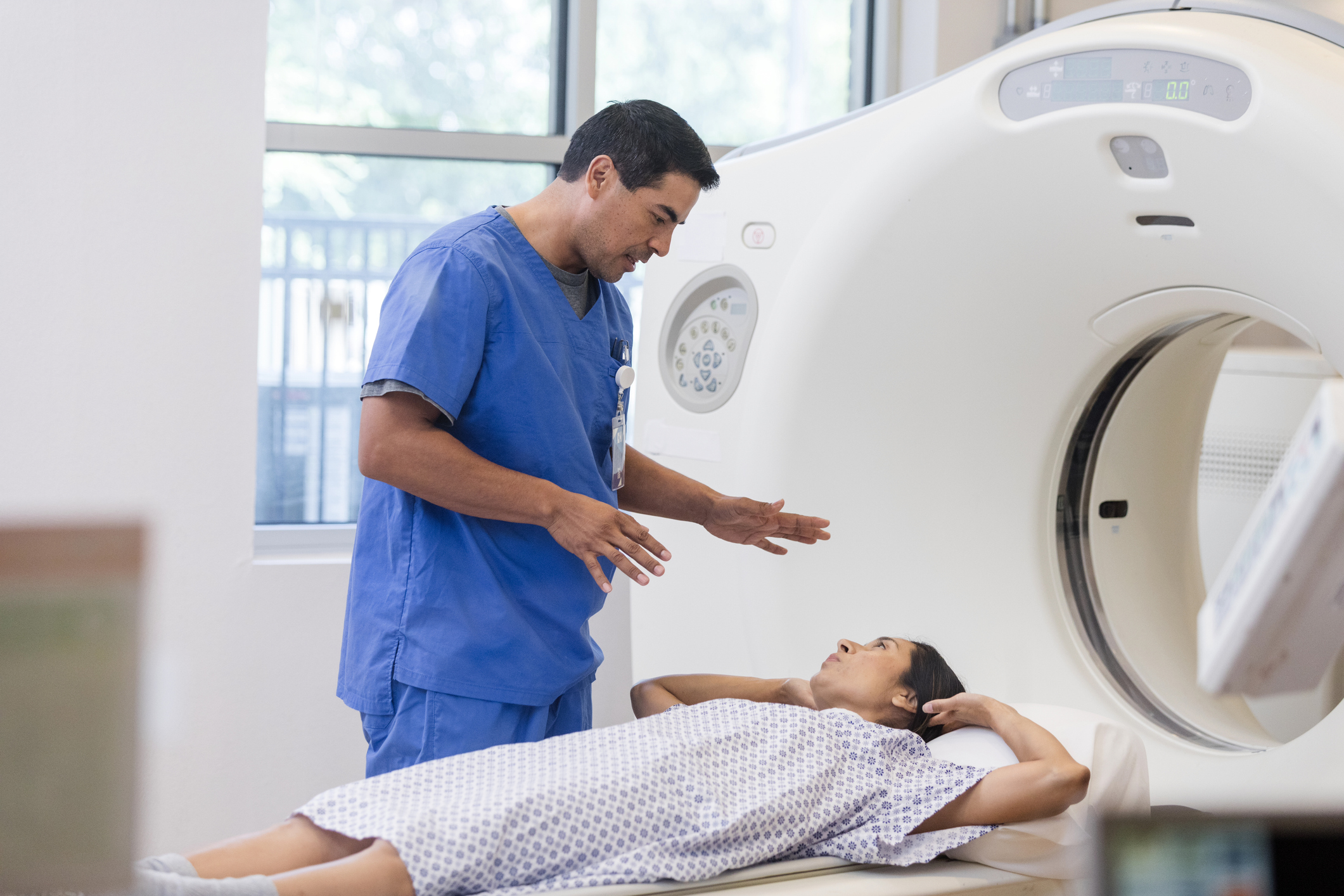
The process starts with a CT scan, which gives a three dimensional picture of the patient’s body, including the tumor to be treated as well as all normal anatomy. This picture may be supplemented with additional information from other 3D images such as PET or MRI scans. Using this picture as a map of the body, the radiation oncologist identifies a target to be treated and any sensitive healthy tissue that needs to be avoided. The radiation oncology team then uses powerful computers to design a radiation plan with multiple beams aimed at the target. Each beam is shaped to deliver the maximum dose possible to the target, while avoiding surrounding sensitive structures. Thus, the radiation “conforms” to the target volume. 3DCRT can be very useful when a tumor is close to a sensitive normal structure. (For example, lung cancer close to the spinal cord.) The added precision of 3DCRT allows the radiation oncologist to give more dose to the tumor while limiting exposure of normal tissue to safe levels.
Since many lung tumors move as a patient breathes, this issue must be taken into account when developing an effective radiation treatment plan. That’s why at Hoag, patients who have tumors that may move with respiration are imaged using state-of-the-art 4-D Computed Tomography (CT).
By utilizing innovative 4-D CT technology, Hoag Radiation Oncologists are able to more accurately take respiratory motion into account when planning radiation therapy. This enables oncologists to more accurately target the cancer and spare healthy tissue during radiation treatments.
Hoag is proud to present Gamma Knife Perfexion, the most advanced and specialized radiation machine for Stereotactic Radiosurgery (SRS). Hoag is the first in Southern California to install the premier unit and continues to be the only facility offering Gamma Knife Radiosurgery (GKRS).
Gamma Knife is the most well studied and published technique for delivering SRS and Perfexion is a revolutionary improvement on the existing technology. As compared to previous Gamma Knife units, the Gamma Knife Perfexion has a cylindrical collimator system that increases the ability to treat lesions independent of their location in the brain. The system also greatly improves patient comfort.
Additionally, the Gamma Knife Perfexion has been configured for maximal shielding outside of the treatment region, which greatly reduces unwanted exposure to the patient. And, the unique collimator system allows for custom configurations, greatly expanding the ability to shape radiation dose to conform to the target volume increasing the capability of sparing larger areas of normal, healthy tissue. The many technological advances of the Gamma Knife Perfexion translate into a safer, more accurate, and more comfortable treatment.
Hoag has a distinct advantage with active SRT/SBRT and SRS programs in the same facility. Our weekly multidisciplinary Neuro-Oncology Tumor Board specialists are therefore able to recommend and carry out optimal treatment regimens within this fully integrated facility assuring the patients the best possible treatment options.
MRI and PET are important tools in the detection and treatment of some cancers. They can detect small differences in the soft tissues of the body which are not seen on CT scans. In Hoag Radiation Oncology, we use these scans in conjunction with the CT scan to more accurately plan patient treatment. This process is referred to as image fusion and allows the physicians and physics staff to identify a patient’s tumor and the normal structures surrounding the tumor with greater precision and sophistication. Hoag’s staff of radiologists are renowned for their work in MRI, PET and MR Spectroscopy and are available for patient consultation with the radiation oncologists as they plan treatment.
The treatment planning system is central to the design of a patient’s radiation therapy plan. Using information gathered from MRI, PET, CT and x-ray images, the treatment planning system creates 3D models of a patient’s tumor and anatomy. Decisions about the how to deliver the radiation to the targeted area can be developed in a virtual reality environment.
The medical dosimetrist will prepare treatment plans and calculate the dose to meet the requirements of the physician’s prescription. The radiation oncologists and medical physicists will then review the treatment plans and approve the most appropriate. The RayStation® Treatment Planning System is among the most sophisticated systems available, with capabilities to create 3D Conformal Radiation Therapy (3DCRT) and Image-Guided Intensity-Modulated Radiation Therapy (IG-IMRT) plans. Software tools such as image fusion and virtual simulation enable a patient’s treatment plan to be developed with the latest technology and greatest precision.
SpaceOAR® hydrogel is the first FDA cleared spacing device to protect the rectum in men undergoing radiation therapy for prostate cancer. Hoag is the first hospital in Southern California, and west of Mississippi, to offer this revolutionary product, and is pleased to offer to patients in both Newport Beach and Irvine.
Because of the close proximity of the prostate to the rectum, prostate radiation therapy typically results in some radiation hitting the rectum, which can sometimes cause side effects. The SpaceOAR System creates space by temporarily pushing the rectum away from the prostate and the high dose area. Placed through a small needle, the hydrogel is administered as a liquid, but quickly solidifies into a soft gel that expands the space between the prostate and rectum. The hydrogel spacer maintains this space until radiation therapy is complete. The spacer then liquefies and is absorbed and cleared from the body in the patient’s urine. This is a significant advancement considering that the most common side effect of radiation therapy is related to proctitis. By creating space between the prostate and rectum, SpaceOAR hydrogel reduces rectum radiation injury, and the resulting long-term complications such as diarrhea, bleeding and pain.
Stereotactic Radiosurgery (SRS) is a technique that delivers a single large dose of radiation to a precisely determined target. It is an alternative or adjunct to neurosurgery or conventional radiation.
The term stereotactic refers to a localizing system that uses a rigid head frame attached to the patient’s skull for precision set-up. Radiosurgery refers to the highly focused beams of radiation that can be used for the same purposes as conventional surgery, but without ever cutting or opening up the patient. The only invasive portion of the procedure is attaching the head frame to the skull, which requires only local numbing medications and light sedation for comfort. There is virtually no recovery time, and the patients go home in the afternoon of their treatment day. In addition, the highly focused beams deliver a larger single dose than possible with other radiation techniques. This large single dose has been found to be more effective in treating certain tumors and conditions.
SRS is most commonly used to treat benign and malignant tumors of the brain, such as brain metastases (cancer that has spread from other parts of the body), meningiomas, pituitary adenomas, acoustic schwannomas, arteriovenous malformations (AVMs), and malignant gliomas (anaplasticastrocytomas and glioblastomamultiforme). SRS is also used to treat some neurologic conditions such as trigeminal neuroalgia, epilepsy, and Parkinson’s Disease.
At Hoag, SRS is performed with the Gamma Knife Perfexion, a highly advanced and specialized radiation machine. SRS performed on the Gamma Knife is also referred to as Gamma Knife Radiosurgery (GKRS).
Hoag has a distinct advantage with active SRT/SBRT and SRS programs in the same facility. Our weekly multidisciplinary Neuro-Oncology Tumor Board specialists are therefore able to recommend and carry out optimal treatment regimens within this fully integrated facility assuring the patients the best possible treatment options.
Radiation therapy is an extremely effective method of treating (non-melanoma) skin cancer. Non-melanoma skin cancer includes cell type diagnosis of basal cell and squamous cell. Superficial (on the skin) treatment for skin cancer (non-melanoma) requires the use of a special machine that will treat the skin but not the underlying tissues. Hoag houses just such a machine. Radiation treatment to skin cancer allows the patient to avoid the alternative option of surgery, which often results in scarring.
By submitting this request, you agree to receive communications from Hoag and accept our Privacy Policy and Terms of Use.