Advancing Breast Health: The Evolution of Breast Imaging
One of the best proactive measures you can take for your breast health is to empower yourself with knowledge. Understand your personal breast health and schedule your mammograms annually.
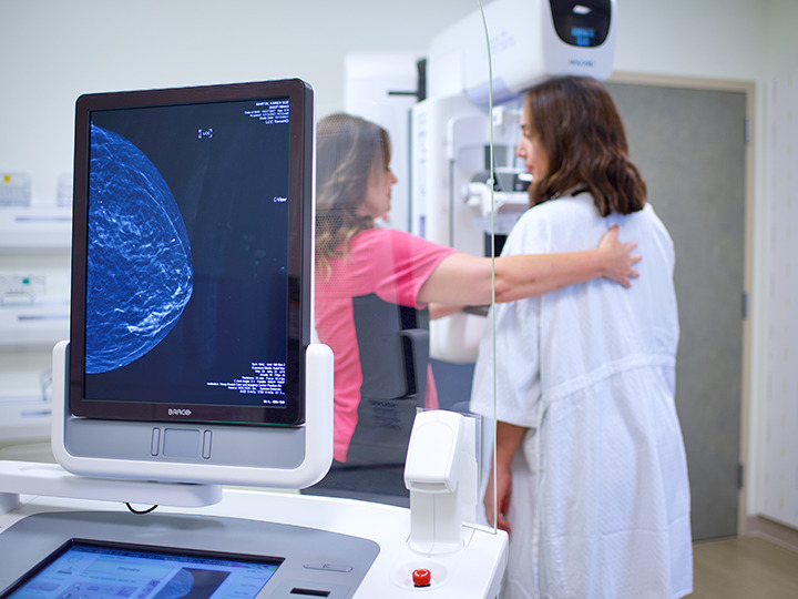
Annual mammograms remain the gold standard for detecting breast cancer. The goal of a screening mammogram is to find cancer at an early stage when treatment is most effective. The National Comprehensive Cancer Network (NCCN), the American College of Radiology (ACR) and the American Society of Breast Surgeons (ASBrS) all recommend yearly mammograms for average risk women starting at age 40, and women at higher risk may be recommended to start even earlier. “Early detection means you have more treatment options. When breast cancer is found and treated early, the national 10-year relative survival rate is greater than 90 percent,” said January Lopez, M.D., medical director, breast imaging at Hoag’s Breast Center.
Our team of fellowship trained breast radiologists are committed to utilizing the latest in advanced breast screening and diagnostic technologies, including standard mammography, tomosynthesis (3D mammography), breast MRI and breast ultrasound, including automated whole breast ultrasound.
3D Mammography or Breast Tomosynthesis: Breast tomosynthesis (3D mammography) was specifically designed to address the limitations of conventional 2D mammography and can be especially beneficial for women with dense breasts. “Breast tomosynthesis can be thought of as a 3D exam since it allows radiologists to examine the breast tissue one layer at a time. Instead of viewing all the complexities of breast tissue in a single flat image, the radiologists can examine the tissue millimeters at a time. Fine details become more visible and are no longer hidden by superimposed breast tissue. With the addition of the newest high-resolution detectors and AI-powered analytics, the quality of 3D mammography is better than ever,” said Dr. Lopez.
Breast Ultrasound: Women with dense breast tissue, breast implants, or changes on their mammogram may benefit from a breast ultrasound, which is a non-invasive, radiation free breast imaging tool that uses high-frequency sound waves to create images of the breast. Ultrasounds can also help shed light on focal breast pain, a lump felt in the breast, nipple discharge or other breast changes.
Traditional hand-held ultrasound is performed by a specially trained breast ultrasound technologist who moves the probe (transducer) over the breast and takes images while scanning. Automated whole breast ultrasound or ABUS uses special ultrasound equipment with a wide transducer that acquires hundreds of images in three or four sweeps across each breast and these images are reconstructed in three dimensions.
“A breast ultrasound does not replace your annual mammogram, but it can be a good complement when additional information is needed,” Dr. Lopez said.
Breast MRI: Magnetic resonance imaging (MRI) is a noninvasive test that uses a magnetic field coupled with an injection of intravenous dye to look at the blood flow patterns of the breasts, which is used along with mammography to detect a certain subset of breast cancers before they are visible on mammography, particularly in women with dense breasts. Mammograms remain essential and can see other subsets of breast cancer earlier than with MRI, but when these two tests are coupled together, they provide the greatest confidence and accuracy in evaluating for breast cancer.
We encourage you to take an active role in your breast health. Hoag offers quick and easy scheduling at seven locations* throughout Orange County. In addition to phone scheduling for all breast imaging, we now offer online scheduling for routine screening mammograms. Visit hoag.org/mammogram to schedule your appointment online.
*Some locations offer walk-in and same day appointments as well as Saturday hours.