
Hoag Hospital Newport Beach
1 Hoag Drive
Newport Beach, CA 92663
(949) 764-5573
1 Hoag Dr, Newport Beach, CA 92663
(949) 764-5573
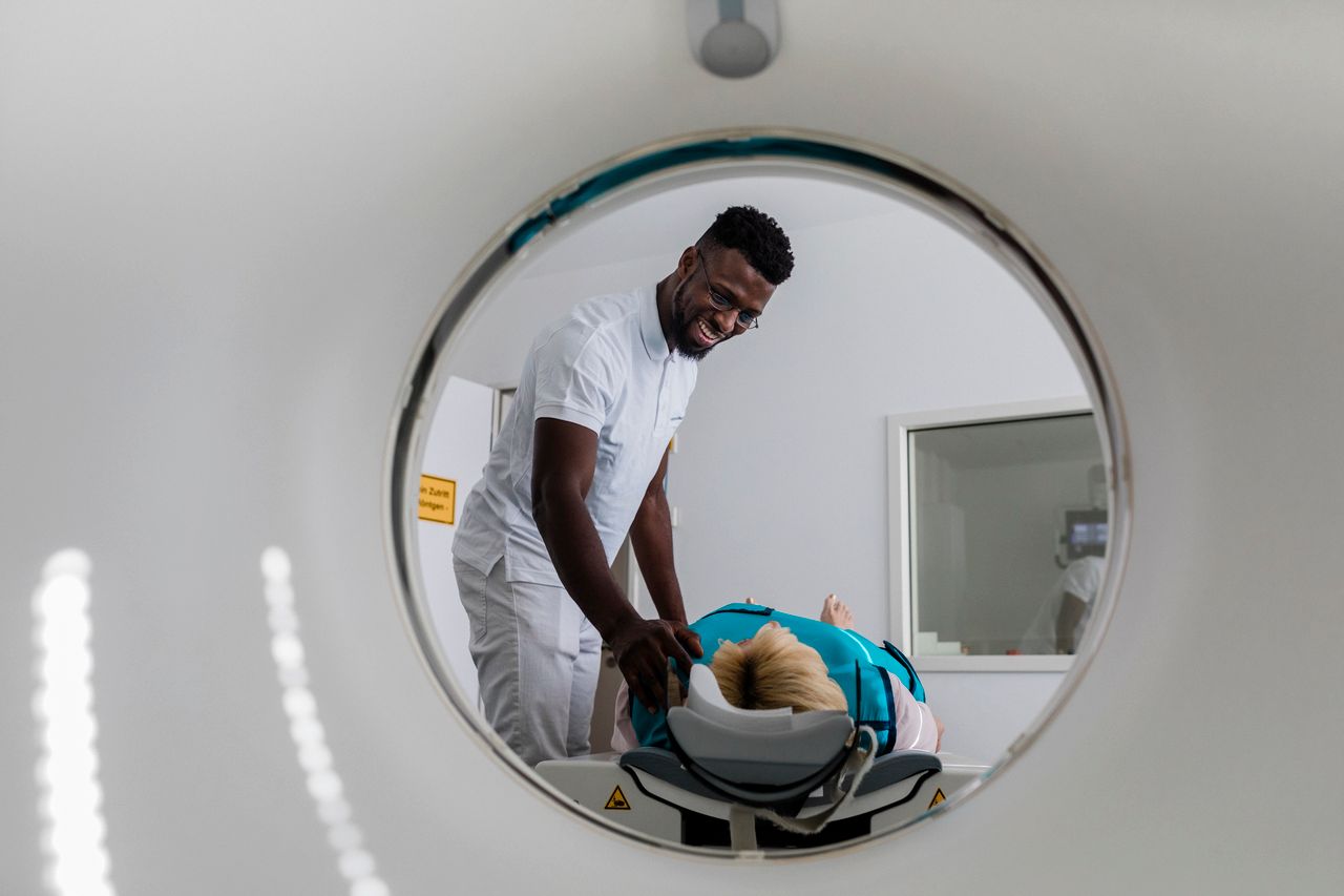

Interventional Radiology

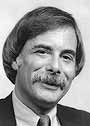
Radiology & Imaging Services Physician, Cardiovascular Radiologist
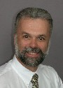
Radiology & Imaging Services Physician
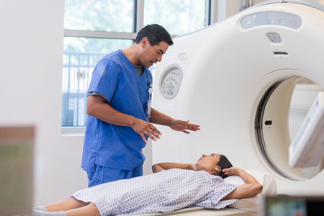
Radiology
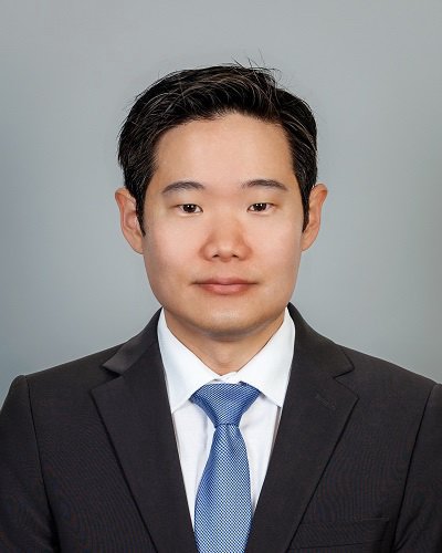
Radiology & Imaging Services Physician

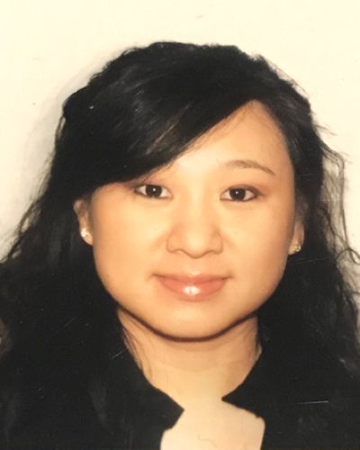
Associate Director, Breast Imaging at Hoag Breast Center
1 Hoag Drive
Newport Beach, CA 92663
(949) 764-5573
26671 Aliso Creek Road, Suite 106
Aliso Viejo, CA 92656
(949) 764-5573
19582 Beach Boulevard, Suite 150 & 160
Huntington Beach, CA 92648
(949) 764-5573
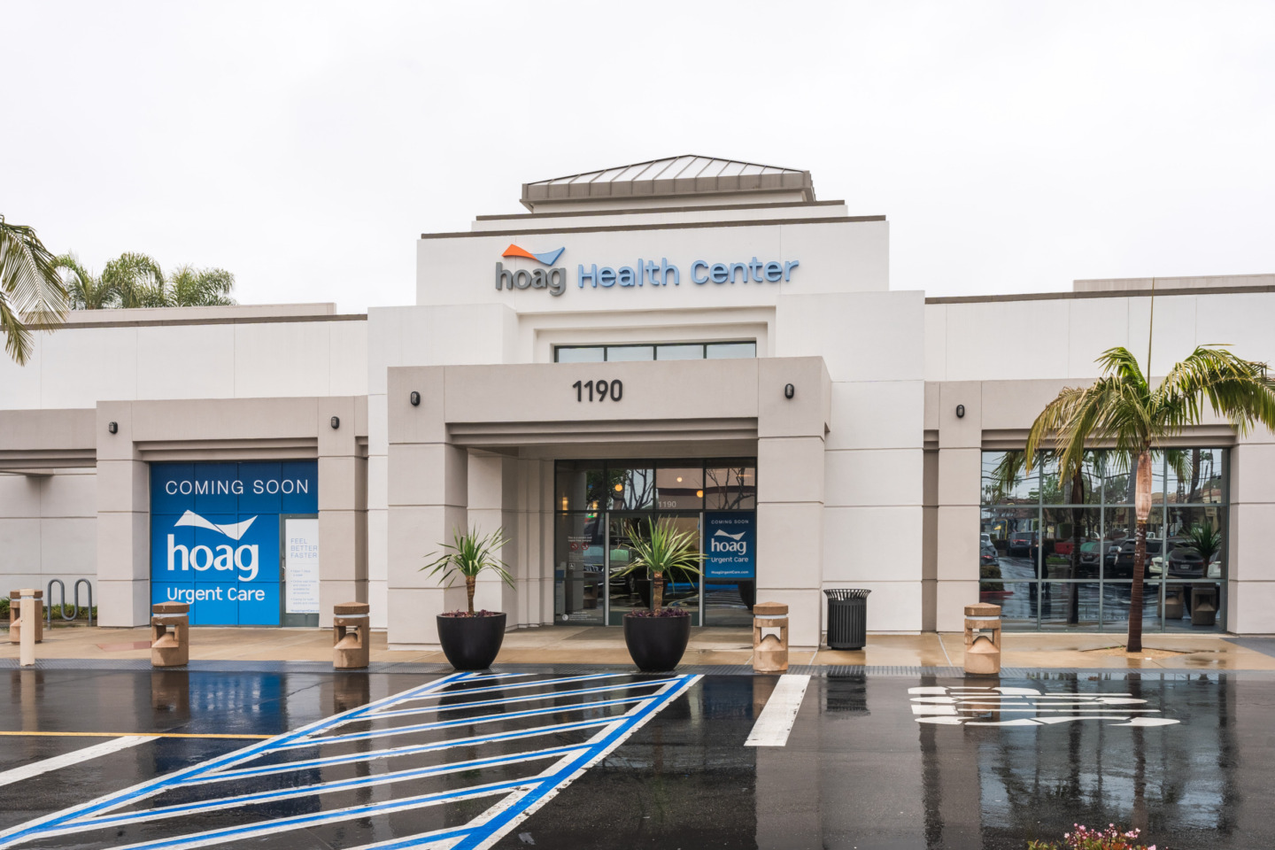
1190 Baker Street, Suite 102
Costa Mesa, CA 92626
(949) 764-5573
16305 Sand Canyon Avenue, Suite 160
Irvine, CA 92618
(949) 557-0180
1 Hoag Drive
Newport Beach, CA 92663
(949) 764-5780
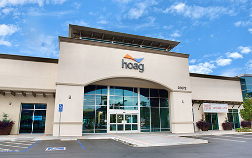
26672 Portola Parkway, Suite 106
Foothill Ranch, Lake Forest, CA, 92610
(949) 764-5573
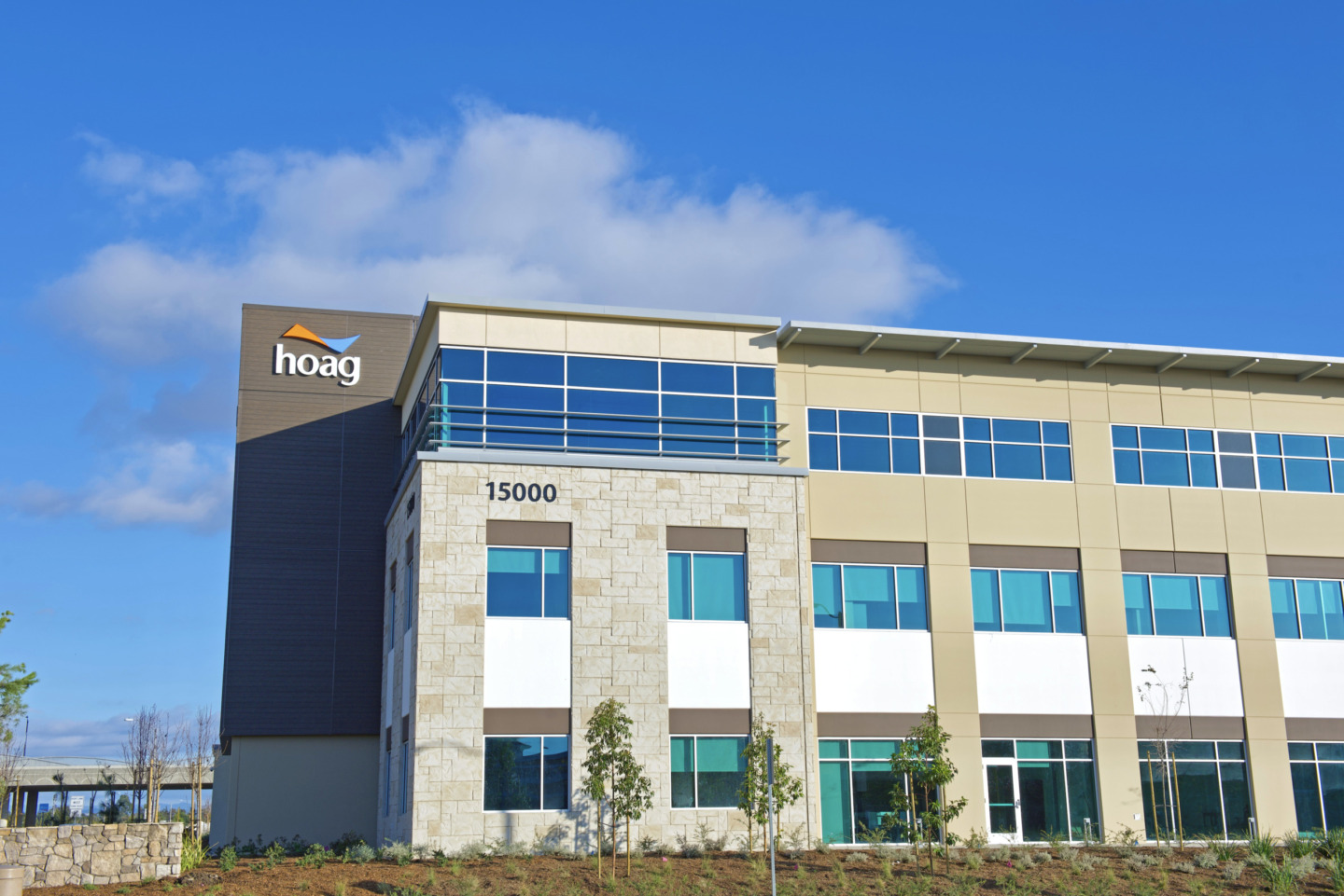
15000 Kensington Park Drive, Suite 150
Tustin, CA 92782
(714) 477-8340
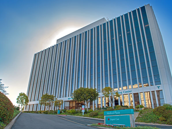
360 San Miguel Drive, Suite 106
Newport Beach, CA 92660
949-721-8191
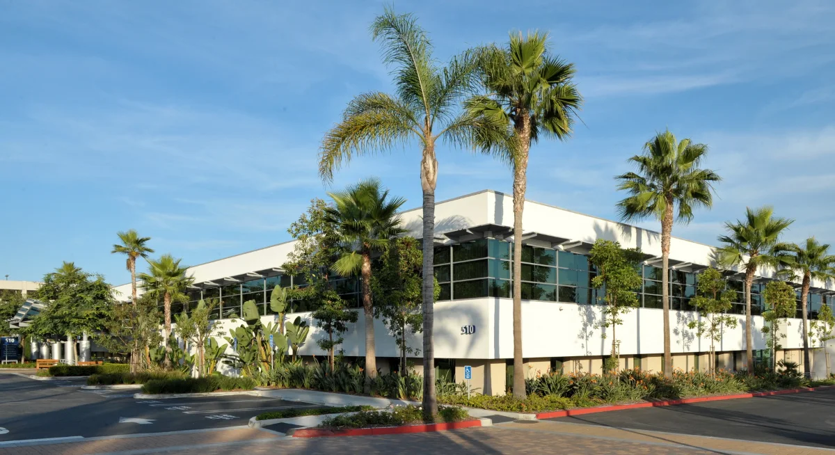
510 Superior Avenue, Suite 100
Newport Beach, CA 92663
(949) 764-5573
1 Hoag Drive, Building #47
Newport Beach, CA 92663
(949) 764-5573
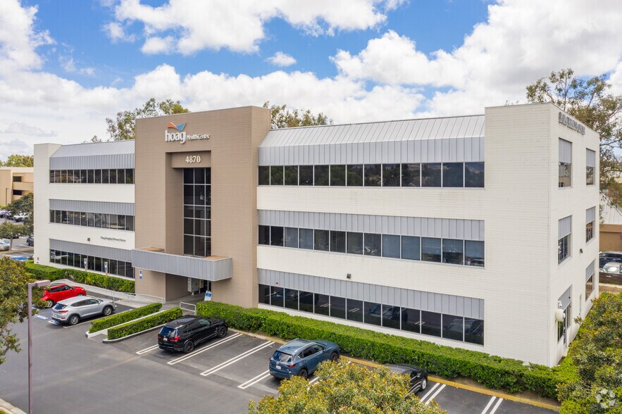
4870 Barranca Parkway, Suite 100
Irvine, CA 92604
(949) 764-5573
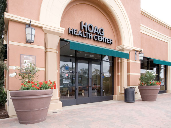
6352 Irvine Boulevard
Irvine, CA 92620
(949) 764-5573
Get care from medical providers that fit your needs in a location near you.
Find a providerDidn’t see what you’re looking for? Reach out and we’ll make sure you get what you need.
Contact usBy submitting this request, you agree to receive communications from Hoag and accept our Privacy Policy and Terms of Use.