Epilepsy Program
520 Superior Ave, Suite 205, Newport Beach, CA 92663
(949) 764-8319
Inquire now
Epilepsy Program
Pickup Family Neurosciences Institute
- About
- Diagnosis
- Treatments
- Epilepsy Monitoring Unit
- Education & Resources
- FAQs
- Meet the Team
Epilepsy Diagnosis at Hoag
Why an Accurate Diagnosis Matters
The first and most important step toward better seizure control is reaching an accurate diagnosis. While this may sound simple, seizures can look very different from one person to another. Some may resemble fainting spells, staring episodes, jerking during sleep, or unusual behaviors. Because many other conditions can mimic epilepsy, it’s essential to see a specialist rather than attempt self-diagnosis.
Our epileptologists will ask detailed questions such as:
When did your seizures begin?
Do you notice any warning signs before a seizure?
What happens to you during an episode?
These answers, combined with advanced diagnostic testing, help us determine whether seizures are epileptic in nature and identify their cause, type, and origin.
Hoag’s epilepsy specialists use a wide range of neurodiagnostic and brain imaging techniques to pinpoint seizure activity.
Advanced Diagnostic Tools
Measures electrical activity in the brain to identify abnormal patterns.
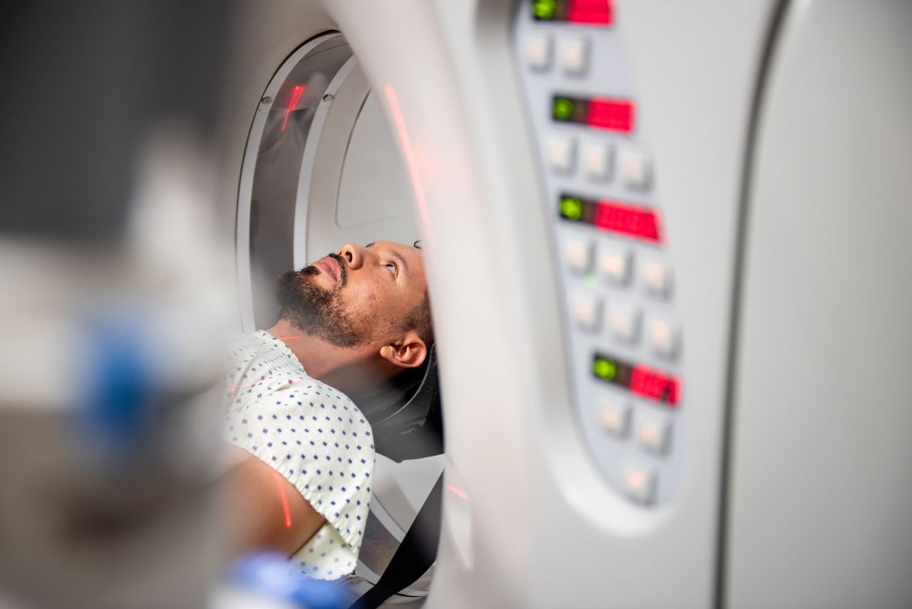
Provides continuous video and EEG monitoring in a safe, comfortable setting to directly observe seizures and determine their source.
For some patients, diagnosis requires observing seizures directly. Hoag’s Level 4 Epilepsy Monitoring Unit offers around-the-clock video and EEG monitoring in a supportive hospital setting. Our team of epileptologists, nurses, and technologists carefully analyzes this data to determine if seizures are epileptic and, if so, their type and origin. This information is critical in designing the most effective treatment plan.
Learn more about Hoag's Epilepsy Monitoring Unit here.
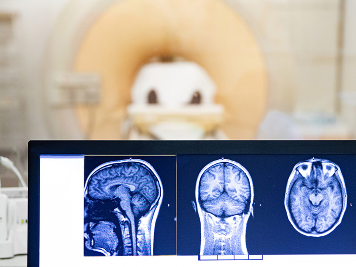
High-resolution 3T MRI scans detect subtle abnormalities. Images may be combined (“fused”) with PET, MEG, or SPECT for even greater detail.
Maps language and motor areas to protect critical brain functions during surgery.
Identifies areas of abnormal brain metabolism linked to seizures
Detects magnetic fields from brain activity, revealing clusters of abnormal signals.
Measures blood flow and metabolism in the brain.
Provides detailed cross-sectional images of the brain, often used in emergencies.
The Bigger Picture: Epilepsy in the US
1 in 26 people will develop epilepsy during their lifetime.
About 3 million U.S. adults live with active epilepsy.
The highest rates are in adults over 60, often due to stroke, tumors, or brain injury.
Two-thirds of patients achieve seizure control with anti-seizure medications.
One-third live with drug-resistant epilepsy, often benefiting from advanced therapies, including surgery.
Epilepsy is linked with other conditions such as autism, multiple sclerosis, cerebral palsy, and depression.
Up to 50,000 deaths annually in the U.S. are related to seizures or complications like SUDEP (Sudden Unexpected Death in Epilepsy).
Epilepsy affects over 3 million Americans of all ages, with nearly 500 new diagnoses daily. Many patients can achieve freedom from seizures or side effects through effective drug therapy or minimally invasive surgical treatments tailored to their needs..
Four Common Questions About Epilepsy
There are a lot of questions that may arise after you have a seizure, but having a seizure doesn't mean that you have epilepsy. Dr. David Millett, Epilepsy Program Director, answers four common questions he receives as an epileptologist.
Level 4 Epilepsy Treatment Center
A Level 4 Epilepsy Center represents the highest standard of care for complex epilepsy. At Hoag’s Pickup Family Neurosciences Institute, earning this distinguished designation means patients benefit from advanced capabilities like inpatient video-EEG monitoring, expert neuroimaging, and access to surgical and neurostimulation treatment options—all delivered by a multidisciplinary team.
Tour the Epilepsy Monitoring Unit
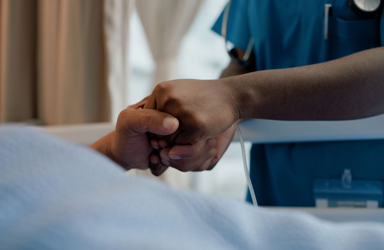
Hoag’s Epilepsy Monitoring Unit provides advanced inpatient care to evaluate and better understand seizures when a diagnosis is unclear or symptoms are difficult to manage. Patients in the EMU are cared for by a dedicated team trained to keep them safe, comfortable, and supported throughout their stay. The EMU is a critical step for many patients on the path to seizure control and improved quality of life.
Seizure Control Means No Seizures
Our goal is simple: to help every person with epilepsy live seizure-free. Even one seizure a year can impact memory, safety, and quality of life, sometimes leading to loss of independence or employment. While medication may help reduce the number of seizures, our epilepsy program is designed to stop them entirely. Through advanced diagnostics, individualized treatment plans, and leading therapeutic options, our team works with each patient to achieve zero seizures and regain control of their life.

Why Choose Hoag?
Common Causes of Seizures

Patient Testimonial Carousel
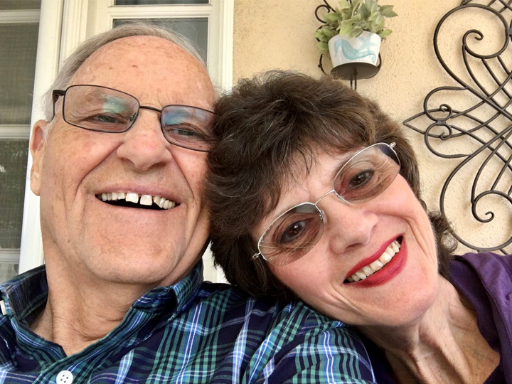
“Through the therapies I received at Hoag, I was slowly able to find my words again, and my memories.”

Helpful Articles
Article
How the Epilepsy Team’s Multidisciplinary Approach Restored a Family’s Smile- October 7, 2025
- 2 min read

Article
After 72 Years of Seizures, Epilepsy Patient Says Hoag Gave Him His Life Back- November 21, 2025
- 3 min read
Your epilepsy care starts here
1
Find the right provider
Get care from medical providers that fit your needs in a location near you.
Find a provider2
Explore epilepsy resources
Find information made to guide and support you on your journey.
Explore Resources3
Get in touch
Didn’t see what you’re looking for? Reach out and we’ll make sure you get what you need.
Contact usMedical Management
At Hoag, our goal is to help every patient live life with fewer seizures and improved quality of life. For many, this begins with medical management—non-surgical treatment options that can effectively reduce or even eliminate seizures. More than 60% of patients achieve good seizure control with antiepileptic medications, making this the most common and effective first step in treatment.
Our epilepsy specialists take into account each patient’s specific diagnosis, health history, and lifestyle when determining the most appropriate therapies. Because medications can sometimes cause side effects or interact with other prescriptions, Hoag’s epileptologists work closely with patients and their primary care physicians to carefully manage drug therapy over time.
More About Medication Treatment for Epilepsy
Antiepileptic drugs (AEDs) are designed to reduce the brain’s tendency to generate abnormal electrical activity that leads to seizures. The goal is not only to stop seizures but also to allow patients to live as safely and independently as possible. For many people, medication is the best way to control seizures while avoiding surgery.
Selecting the right medication is a personalized process. Hoag specialists will:
Review seizure type and frequency:
Different medications work better for different seizure types.
Assess patient lifestyle and health:
Factors such as age, pregnancy plans, and other health conditions guide treatment choices.
Start with the lowest effective dose:
To minimize side effects while achieving seizure control.
Adjust over time:
Medications are often fine-tuned based on response and tolerance.
Because epilepsy medications can interact with other drugs, it’s essential to have ongoing management from an epileptologist—a neurologist with specialized training in seizure disorders.
While treatment is always individualized, commonly prescribed antiepileptic drugs may include:
Levetiracetam (Keppra®)
Often well tolerated with fewer drug interactions.
Lamotrigine (Lamictal®)
Frequently used for focal and generalized seizures, with mood-stabilizing benefits.
Valproic acid (Depakote®)
Effective for many seizure types but not recommended in all patient populations.
Topiramate (Topamax®)
May be used in patients with both epilepsy and migraine.
Your care team will discuss the risks, benefits, and alternatives to ensure you understand the best option for your situation.
The Importance of Seeing an Epileptologist
While primary care physicians and general neurologists play an important role, epilepsy care is most effective under the guidance of a specialist trained in seizure disorders. Epileptologists at Hoag:
Provide precise diagnoses based on seizure type and underlying causes.
Help patients navigate complex medication decisions.
Manage side effects and long-term health impacts.
Explore advanced therapies if medications alone are not successful.
With expert guidance, patients can feel confident they are receiving the most up-to-date, effective care available.
Surgical Treatments
In approximately one third of patients, anticonvulsant medications cannot adequately control seizures. When seizures cannot be controlled with medication alone, Hoag offers the latest techniques in surgical intervention. Surgery can eliminate seizures in the majority of patients and reduce seizures in most of the remainder. Various testing is done to help determine where seizures are starting in the brain. This may include implantation of electrodes for intracranial monitoring. Once the location causing seizures has been determined, Hoag neurosurgeons utilize state-of-the-art neuronavigational technology and sophisticated magnetic resonance imaging techniques to precisely reach and treat the seizure areas safely.
Types of Surgical Treatments Available at Hoag
Stereoelectroencephalography (sEEG) is a diagnostic procedure using miniature electrodes measuring less than 1 mm in diameter to record brain activity in the location of seizures. This minimally invasive technique does not require traditional incisions or hair shave, while still permit a 3-dimentional map of the seizure network. By localizing seizures with sEEG, Hoag neurologists and neurosurgeons can predict how to cure seizures with subsequent treatments. Patients stay in the Epilepsy Monitoring Unit for one week, on average, during brain mapping and go home the day after sEEG removal.
Laser therapy is a minimally-invasive, hair-sparing procedure that can cure seizures by treating the seizure-origin within the brain. Following a short procedure performed in the MRI scanner, patients stay overnight before returning home. Since there is no incision, patients return to normal activities quickly with minimal discomfort.
Patients who have benefited from this technology have a 87% seizure freedom rate compared to the national average of 61%.
MR-guided-focused ultrasound is a technique used to treat the brain noninvasively. This treatment is FDA-approved for movement disorders and used on clinical trials to treat seizure disorders without requiring general anesthesia or operation.
RNS technology combines real-time surveillance for seizures with gentle stimulation to stop seizures at their origin before they spread. This fully implantable neuromodulation system reduces seizures by up to 80%. Importantly, RNS is the only technology that records seizure frequency and EEG at home. This provides valuable information for the patient’s provider when managing treatment.
Originally developed in the 1980’s, this technology gained approval for the treatment of seizures in recent years. DBS blocks seizure propagation, preventing seizure spread even when seizures are generalized. Seizures may be reduced by up to 80%. Additionally, the system is rechargeable, thereby tripling the life expectancy of the implanted battery before needing replacement.
VNS activates the vagal nerve in the neck to modulate nervous system activity. VNS is an effective therapy for those who are not candidates for other therapies. VNS may reduce seizures by 50-60% and be used in children as young as four years old.
Hoag offers minimally-invasive approaches to reduce or cure seizures. These techniques include noninvasive procedures, minimally-invasive procedures, and endoscopic procedures. By reducing trauma to the tissue, patients are exposed to less surgical risk, faster recovery, less pain, and improved cosmetic result.
Hoag utilizes hair-sparing surgical approaches. From laser surgery to open surgery, our neurosurgeons utilize plastic-surgery style approaches to maximize positive results. In many of our procedures, minimal- or no-hair is shaved during surgery. This permits individuals to return to normal activities without the feeling of stigma associated with brain surgery.
Hoag surgeons have mastered surgical approaches by training across the globe to bring the latest surgical techniques to the individuals of Orange County. The range of surgical approaches routinely performed at Hoag include craniotomy, lobectomy, lesionectomy, topectomy, corpus callostomy and disconnection procedures.
The Pickup Family Neurosciences Institute at Hoag became one of the first hospitals in the nation to obtain and use the Medtronic Stealth Autoguide Robotic Platform to treat epileptic seizures and the first hospital in the world to perform minimally invasive brain biopsy using this technology.
The Stealth Autoguide Robot uses a small pinpoint hole through which neurosurgeons can place electrodes or biopsy needles precisely in the brain to diagnose or treat epilepsy, cancer or other neurological conditions. Without this tool, these procedures typically involve shaving large portions of the scalp, making larger incisions and removing pieces of skull to expose the brain for surgery.
The highly advanced surgical tool replaces open cranial surgery for certain patients and greatly increases accuracy and patient recovery. The device can be used for a range of neurological conditions that require precision guidance for placement of diagnostic electrodes, biopsy or targeted laser ablation.
About the Epilepsy Monitoring Unit
For some patients with epilepsy or unexplained seizures, traditional tests such as MRI or routine EEG may not provide enough information to confirm a diagnosis or guide treatment. In these cases, your neurologist may recommend a stay in Hoag’s Epilepsy Monitoring Unit (EMU), a specialized inpatient program designed to capture and study seizure activity in a safe, controlled environment.
Hoag's EMU is designed with patient comfort in mind. Patients stay in private rooms that are not only equipped with the latest medical monitoring technology but also feature scenic views to enhance the healing environment. These comfortable rooms allow patients to feel relaxed during their stay, while knowing they are under constant medical supervision for safety.
Why You May Be Offered EMU Care
You may be referred to the EMU if:
Your seizures are difficult to diagnose.
Your medications are not controlling your seizures.
You may be a candidate for surgical or device-based therapies.
Your care team needs more detailed information to tailor your treatment plan.
By recording seizures as they occur, EMU specialists can determine the type, frequency, and origin of seizures, leading to more accurate diagnosis and targeted treatment.
One of the primary goals of Hoag’s EMU is to accurately classify seizures, which is crucial for determining the most effective treatment. Many patients who have been misdiagnosed or have complex seizure types find clarity at Hoag, where the EMU team performs thorough evaluations to pinpoint the exact nature of their condition. For patients with epilepsy, the data collected helps the team refine their treatment approach, whether through adjusting medications, recommending lifestyle changes, or considering surgical interventions.
Results You Can Expect
During your EMU stay, continuous monitoring provides insights that are often not possible in an outpatient setting. Outcomes may include:
Precise diagnosis of seizure type and whether events are epileptic or non-epileptic.
Optimization of medication therapy, ensuring the right drug at the right dose.
Evaluation for advanced therapies, such as surgery, neurostimulation devices (e.g., RNS, VNS, DBS), or dietary interventions.
Improved quality of life through better seizure control and reduced side effects.
Your Care Team
At Hoag’s Level 4 Epilepsy Center, EMU patients are cared for by a multidisciplinary team, including:
Neurologists
Epileptologists (neurologists with advanced training in epilepsy)
Specialized nurses trained in seizure care and safety
Neurodiagnostic technologists who monitor EEG data 24/7
Neurosurgeons, neuropsychologists, and radiologists who collaborate on complex cases
Pharmacists and dietitians who provide medication and dietary support when appropriate
This team-based approach ensures that every aspect of your care is coordinated and personalized.
Why Choose Hoag's EMU?
Hoag’s EMU is part of our comprehensive Level 4 Epilepsy Center, the highest designation awarded by the National Association of Epilepsy Centers. This recognition reflects Hoag’s expertise in:
Complex diagnostic testing
Advanced surgical and non-surgical therapies
Multidisciplinary care and collaboration
Access to clinical research and trials for patients with drug-resistant epilepsy
With state-of-the-art monitoring technology and one of the most experienced epilepsy teams in Southern California, Hoag provides patients and families with answers, options, and hope.
First Aid for Epilepsy
If you see someone having a seizure, follow this guide to keep the person safe until the seizures stop naturally and full awareness returns. If the person falls to the ground, assist them in keeping their head away from harm and injury.
Do
Keep calm
Look at a clock and note the time of the seizure
Clear the area around the person of anything hard or sharp
Loosen ties or anything around the neck that may make breathing difficult.
Put something flat and soft, like a folded jacket, under the head. Stay with the person until the seizure ends naturally.
After the seizure, place the person on their side. This will help keep the airway clear.
Be reassuring and use a calm voice as consciousness returns.
Do Not
Do not hold the person down or try to stop their movements.
Do not try to force the mouth open with any hard implement or with fingers. A person having a seizure CANNOT swallow their tongue. Efforts to hold the tongue down can injure teeth, jaw, or your own fingers.
Do not attempt artificial respiration except in the unlikely event that a person does not start breathing again after the seizure has stopped.
First Aid for Partial and Non-Convulsive Seizures
You don’t have to do anything if a person has brief periods of staring or shaking of the limbs. If someone has the kind of seizure that produces a dazed state and automatic behavior, the best thing to do is:
Speak quietly and calmly in a friendly way.
Guide the person gently away from any danger, such as a steep flight of steps, a busy highway, or a hot stove.
Don’t grab hold of the person unless some immediate danger threatens. People having this kind of seizure are on “automatic pilot” as far as their movements are concerned. Instinct may make them struggle or lash out at the person who is trying to hold them.
Stay with the person until full consciousness returns, and offer help as needed.
Confusion may occur during a complex partial seizure or during the recovery period after other types of seizures. If the confusion persists, seek medical attention.
Frequently Asked Questions
Hoag is a Level 4 Epilepsy Center, the highest accreditation for epilepsy care. Our team includes three full-time epileptologists, a functional neurosurgeon, and specialized nurses and technologists. We combine advanced diagnostics, personalized treatment, and compassionate care to help patients achieve the best possible outcomes.
The first step is a detailed discussion with one of our epilepsy specialists (epileptologists). Your doctor will ask questions about your seizure history, symptoms, and any warning signs you may notice before an episode. This information helps us decide which tests are needed to confirm a diagnosis.
Our specialists use a variety of advanced tools, including:
Electroencephalogram (EEG): Records brain activity.
MRI and fMRI scans: Provide detailed images of the brain.
PET, MEG, and SPECT scans: Show brain function and metabolism.
Epilepsy Monitoring Unit (EMU): Allows doctors to safely observe and record seizures
An EMU is a specialized hospital unit where patients stay under continuous video and EEG monitoring. This allows doctors to see exactly what happens during a seizure, determine whether seizures are epileptic, and identify where in the brain they start.
There are many things that can cause increased risk of seizures in people that are prone to seizures. People with epilepsy should not drink alcoholic beverages or do illicit drugs. Doing so will increase the risk of seizures significantly. Many people with epilepsy have identified certain things that seem to increase the number or severity of their seizures. Sometimes these connections are just coincidences, but in many cases a link has been proven between these factors (also called triggers) and the occurrence of seizures.
Examples of typical seizure triggers include: lack of sleep, skipping a meal, or increased stress.
Many conditions can mimic epilepsy, such as fainting, sleep disorders, or movement disorders. An accurate diagnosis ensures you receive the right treatment and avoid unnecessary medications.
About two-thirds of people with epilepsy achieve good seizure control with medications. For the one-third of patients with drug-resistant epilepsy, advanced options such as surgery, neurostimulation, or dietary therapy may be recommended.
People who have recently had a seizure or are having active seizures should not drive. However, people with epilepsy whose seizures are fully controlled with medication can qualify to drive. With close follow-up with a neurologist, you can often drive after being seizure-free for a period of time. The physician will work with you to help you drive as soon as your seizures are controlled and there is no risk of losing consciousness from seizures.
If driving is not an option, alternatives such as using public transportation, signing-up with local services for the elderly or disabled, or even moving to an apartment complex or community that has its own transportation may be among the alternatives.
Women with seizures and on seizure medication can get pregnant. However, certain medications can cause harm to an unborn child. There are many medications that are safe and supplements can be taken to decrease the risk of certain birth complications associated with epilepsy medication. The neurologist should be made aware of any upcoming planned pregnancy. The doctor will work with your obstetrician to provide individualized counseling or make changes to current medications.
Meet The Team

David E. Millett, MD
Program Co-Director, Epilepsy
James Park, DO
Program Co-Director, Epilepsy
Timothy H. Lucas Jr, MD
Chief of Neurosurgery
Joshua J. Martin, MD
Epileptologist

Andrew D. Ly, MD
Concussion/Mild TBI Program Program Advisor, Neurophysiology Services

Jason S. Muir, MD
Program Director, Neurophysiology Services, Board Certified Sleep Specialist, Staff Neurologist

Lauren L. Bennett, PhD
Clinical Neuropsychologist

Ruth T. Morin, PhD
Clinical Neuropsychology
Stephanie Chow, MSN, RN, CCRN
Epilepsy Program Nurse Navigator
Stay up-to-date on the latest news from Hoag
By submitting this request, you agree to receive communications from Hoag and accept our Privacy Policy and Terms of Use.