How breast imaging is evolving to support more personalized care
"If mammograms have worked for decades, why do I keep hearing about other types of imaging scans?" I hear this question every time a new technology grabs the headline, and the question is a fair one.
While mammography remains the gold standard for breast cancer screening, the evolution of imaging technology is expanding our ability to create a truly personalized approach to care based on each woman’s actual risk. Personalization will make cancer prevention, detection and treatment more powerful and give women more agency over their health. But standard mammography will not—and should not—be going away any time soon.
Since standard mammogram screening became the norm in the 1970’s, the breast cancer death rate has fallen by 40 percent. Today, advanced imaging is improving our ability to serve more people with different risk profiles better and more effectively. A 45-year-old with dense breast tissue and a strong family history needs a different plan than her best friend who has neither. Advanced breast imaging technologies allow doctors to create the best plans for both.
Three-dimensional tomosynthesis enables radiologists to scroll through breast tissue in thin slices instead of one flattened image. This makes it easier to spot abnormalities in dense breast tissue, for which standard mammography's sensitivity can drop as low as 30 to 48 percent. The difference this has made is so significant that federal law now requires every mammogram report to disclose a patient’s breast density.
Although some cancers are only visible on mammograms, for women with dense breast tissue, we add supplemental tools, such as breast ultrasound, to help detect cancers that can be masked by dense tissue. And for those with a strong family history or a known mutation like BRCA1 or BRCA2, breast MRI, which looks at blood flow patterns to detect certain types of cancer before they show up on a mammogram or ultrasound, is a powerful tool.
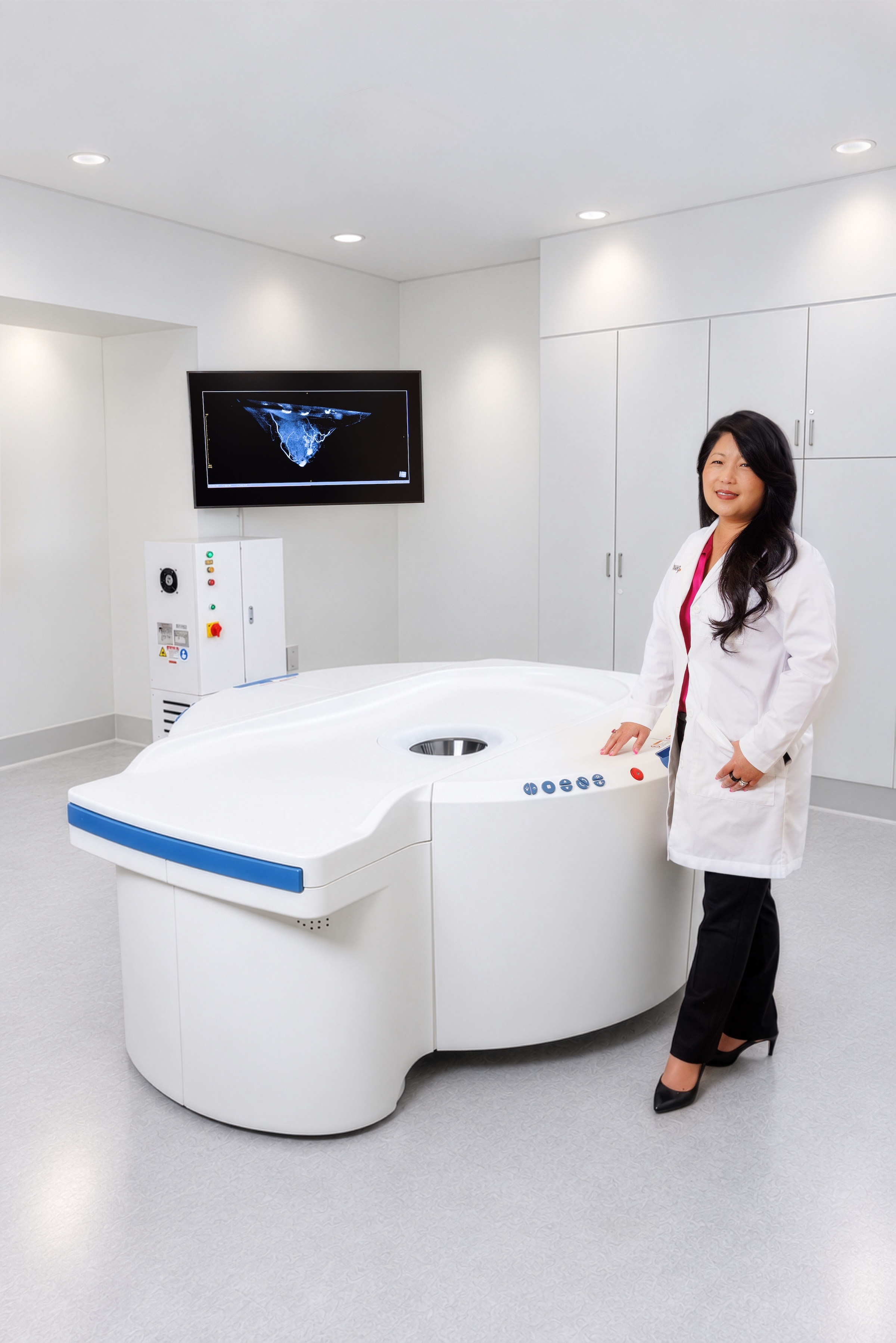
Innovative breast care centers are also embracing groundbreaking technology that is reshaping the way breast cancer is detected. Hoag is the first hospital in Orange County to offer the Koning Vera Breast CT, an advanced imaging technology that delivers high-resolution, 3D images that enable accurate and early detection of breast cancer without breast compression and with minimal radiation. Early studies of contrast-enhanced breast CT have shown high sensitivity for detecting breast cancer, including cancers that may be difficult to see on conventional mammography. Researchers are continuing to evaluate its role in detecting and diagnosing breast cancer, but its ability to provide detailed, three-dimensional images without compression represents an important step forward in breast imaging.
Artificial intelligence is also opening new possibilities in breast imaging. Studies have shown that AI may augment and even surpass our current ability to predict a person’s long-term risk of breast cancer. Think of it like your radiologists’ assistant, another set of eyes to help identify people who might benefit from additional screening or testing while minimizing unnecessary testing for others. Researchers are also beginning to explore AI applications in newer technologies such as breast CT.
All these tools point to a future in which women can expect more personalized prevention plans—and fewer anxiety-producing callback appointments and unnecessary procedures. Personalized breast imaging means a woman and her doctor look at her real risk factors, family history, breast density and genetics, and decide together which combination makes sense for her. For one patient, that may be an annual mammogram. For another, it could mean a mammogram plus an annual MRI. Every woman is different, every cancer journey is different, and technology helps match the right plan to the right patient.
The mammogram you’ve been getting is not being replaced or made obsolete. It’s being supported by a growing set of tools, and by physicians using them to tailor your screening specifically to your risk profile, your lifestyle and your goals.
Dr. January Lopez is a board-certified diagnostic radiologist and fellowship-trained breast imaging subspecialist at Hoag Memorial Hospital Presbyterian in Newport Beach, Calif.
Related Articles

Article
How team-based endometriosis care restored a woman’s quality of life- October 12, 2026 · 4 min read