Skull Base & Pituitary Tumor Program
3900 Pacific Coast Hwy, Newport Beach, CA 92663
(949) 764-4624
- About
- Conditions Treated
- About Skull Base Surgery
- Treatments
- Meet the Team
Skull Base Surgery
Skull base neurosurgery is a specialized surgical approach focused on the area at the bottom of the skull, which includes complex anatomical structures like nerves, blood vessels, and the brain. This type of surgery is often employed to remove tumors, repair abnormalities, or address conditions affecting the base of the skull and surrounding areas. It requires a high level of expertise due to the intricate nature of the region and the proximity to critical brain structures.
Techniques used in these surgeries can be either open or minimally invasive. Open surgeries involve making an incision to access the skull base, while minimally invasive techniques use endoscopes through the nose or small openings. The choice of technique depends on the location and type of the condition. The surgery aims to remove or repair the problematic area while minimizing damage to nearby critical structures like the brain, cranial nerves, and blood vessels. Advanced imaging and navigation technologies often assist surgeons in achieving precise results.
Virtual & Augmented Reality in Neurosurgery
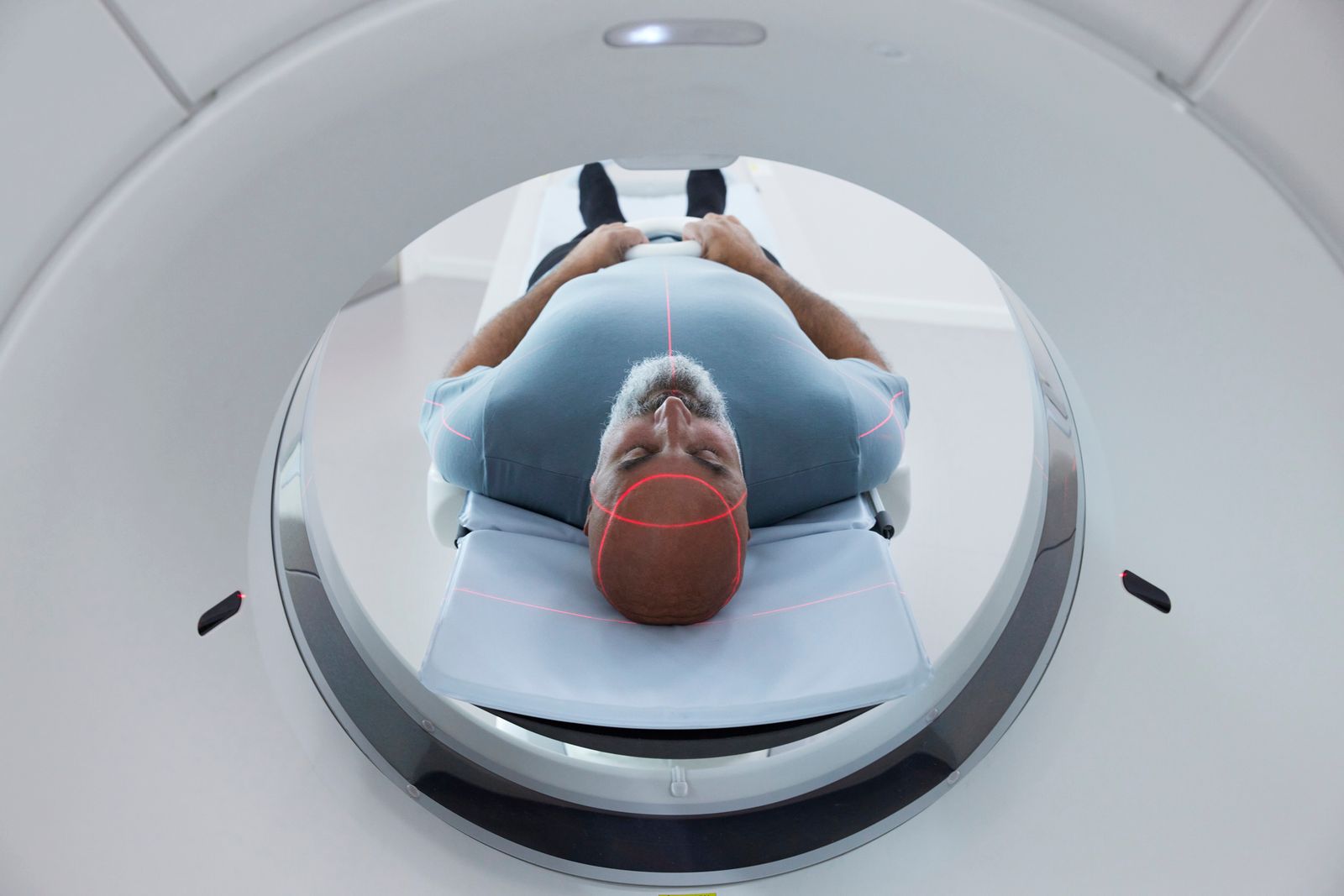
At Hoag, advanced technology meets surgical artistry. Using the Precision Virtual Reality® platform by Surgical Theater, our neurosurgeons transform traditional MRI and CT scans into an immersive 3D model of your brain. This allows our experts to study every angle of your anatomy and rehearse the procedure virtually before entering the operating room.
Patients and their families are invited to “walk the surgery” alongside their surgeon, gaining a clear understanding of where the tumor is located and how it will be safely removed. This approach not only enhances precision but also eases anxiety, turning fear of the unknown into confidence and collaboration.
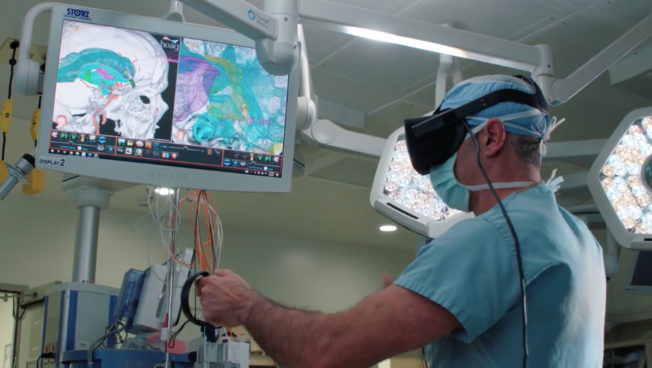
During surgery, the same technology transitions into an augmented reality (AR) experience, overlaying the surgical path within the surgeon’s microscope. It’s like having X-ray vision, offering unparalleled accuracy and minimizing risk to surrounding tissue.
Hoag remains one of the nation’s highest-volume centers using AR in neurosurgery and continues to advance the field under the leadership of Dr. Louis, Empower360 Endowed Chair in Skull Base and Minimally Invasive Neurosurgery.
Inside the Surgical Theater
The Skull Base and Pituitary Tumor team uses the patient's MRI images to create a virtual map of the brain. Using this technique allows the neurosurgeon to see exactly where the tumor lies and the surrounding structures that may be impacted during surgery. Once the full view of the skull and its structures has been made into the model, the surgeon can map out the surgical path that will allow for the least complications and best outcomes. This is also when the patient and their families can view the surgical path and discuss with their surgeon any questions they may have.
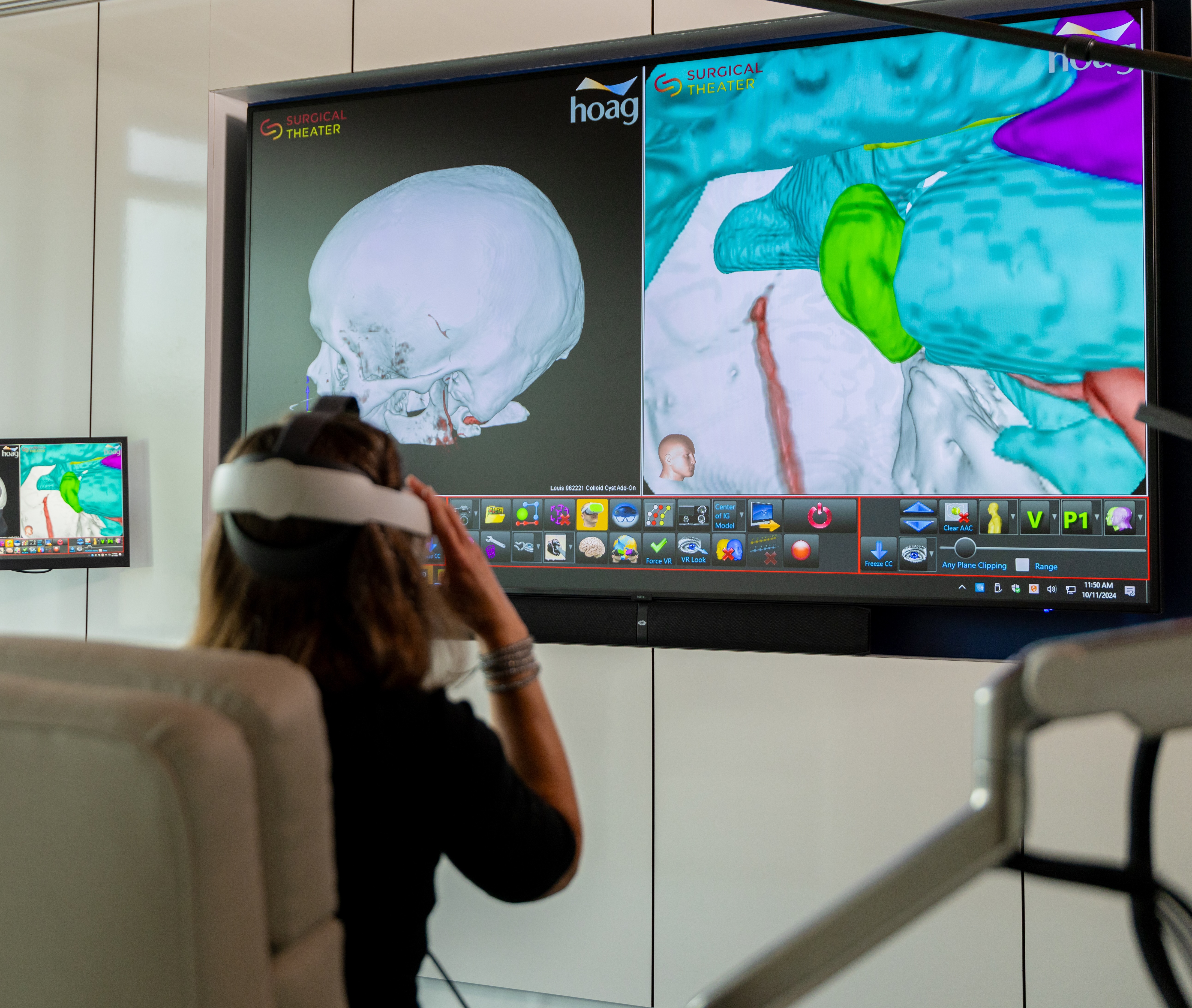
Pre-surgery Rehearsal
After the surgical path has been confirmed, the neurosurgeon can then "practice" the surgery in a virtual format. This allows the surgeon to know exactly where the structures lie, where they can enter the brain and what obstructions they may encounter. Practicing in a virtual environment allows the surgeon to become comfortable with the unique structure and allows for consistency in approach.

Inside the OR
After mapping the brain, reviewing the surgical pathways and rehearsing the surgery, the neurosurgeon is ready to begin removal of the obstruction. The same image that was used in the virtual reality setting is now overlaid in the microscope. This augmented reality allows the surgeon to have "X-ray vision" to see the structures before and during entry. This method allows for precision and best outcomes for the patient.


Stay up-to-date on the latest news from Hoag
By submitting this request, you agree to receive communications from Hoag and accept our Privacy Policy and Terms of Use.