- About
- Diagnosis
- Treatment
- Meet the Team
Types of Thyroid Cancer
Thyroid cancer is the most common type of endocrine cancer. Although a diagnosis of cancer is always concerning, the vast majority of thyroid cancers are very treatable and are associated with an excellent prognosis.
There are four main types of cancer of the thyroid:
Papillary thyroid cancer is the most common, comprising about 80% of all thyroid cancers. It tends to grow slowly but may spread to lymph nodes in the neck or elsewhere in the body. With early intervention, however, papillary thyroid cancer generally has an excellent prognosis.
Follicular thyroid cancers represent about 15% of all thyroid cancers. Follicular thyroid cancers usually do not spread to the lymph nodes, however, in some cases they can spread to other parts of the body, such as the lungs or bones.
Medullary thyroid cancer (MTC) represents about 3% of all thyroid cancers. There are two types of medullary thyroid cancer: sporadic and familial. Approximately 35% of all MTC runs in families and may be associated with other endocrine tumors. Genetic testing (of the RET proto-oncogene) is recommended for those newly diagnosed with MTC. For individuals with a family history, it is helpful to determine whether there are genetic markers present. In individuals with these genetic changes, prophylactic surgery has a high probability of being curative.
Anaplastic thyroid cancer is difficult to control and treat because it is a very aggressive type of thyroid cancer. Anaplastic thyroid cancer is quite rare, making up less than 2% of patients with thyroid cancer
Risk Factors and Causes of Thyroid Cancer
There are several risk factors that can increase an individual’s chances of developing thyroid cancer, such as a family history of thyroid cancer, gender (women have a higher incidence of thyroid cancer), age (the majority of cases occur in individuals over age 40, although thyroid cancer can affect all ages), and history of ionizing radiation exposure. If you have a family history of thyroid cancer, or other risk factors, speak with your physician about whether thyroid screening and genetic testing may be appropriate for you.
Diagnosing Thyroid Cancer
Diagnosing thyroid cancer in its earliest stages can increase the probability of your treatment being more successful. Hoag’s thyroid cancer team is highly skilled in diagnosing and staging thyroid tumors using the latest in state-of-the-art imaging, ultrasound-guide needle biopsy and other specialized tests, including advanced nuclear medicine studies. Upon analysis of test results, Hoag’s multidisciplinary thyroid cancer team develops a personalized treatment plan that addresses all facets of care.
Blood Tests
There are several types of blood tests that may be utilized to diagnose and monitor thyroid cancer patients during and after treatment. Tests for thyroid management include: thyroid hormone levels, thyroid stimulating hormone (TSH) and thyroglobulin. Other blood testing involving molecular markers may also be used, as well as genetic testing for certain types of thyroid cancer.
Imaging Tests
There are several types of imaging studies that may be utilized during diagnosis and to monitor thyroid cancer patients during and after treatment. The most common imaging tests used for diagnosing thyroid cancer include:
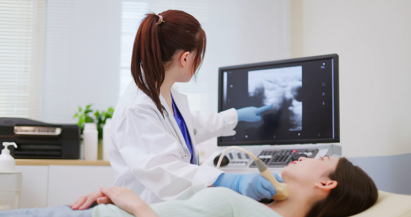
Ultrasound. Ultrasound is an imaging study that uses high-frequency sound waves to create pictures of internal organs. This non-invasive test can help physicians determine the number and size of nodules on the thyroid. It can also help determine whether a nodule is solid, filled with fluid (cyst) or complex (mixed solid and fluid). Ultrasound is an excellent modality for evaluation of the lymph nodes in the neck for possible involvement with thyroid cancer.
Computerized Tomography (CT). A CT scan is procedure that uses a computer to produce three-dimensional, cross-sectional images of inside the body. CT scans are sometimes ordered for patients with thyroid cancer to examine parts of the neck that cannot be optimally visualized with ultrasound, as well as to determine if the cancer has spread to other areas of the body.
Magnetic Resonance Imaging (MRI). MRI produces images of the body’s internal structures by passing radio waves through a powerful magnetic field. Differing frequencies of radio waves are produced by the different body structures. In return, these are mapped and converted into digital images by a computer. MRI helps clinicians to distinguish between normal and diseased tissue to identify cancerous cells within the body, and is also useful for exposing metastases. MRI provides greater contrast within soft body tissues as compared to a CT scan.
Laryngoscopy. Because the thyroid gland is so close in proximity to the vocal cords, thyroid tumors may sometimes affect them. During a laryngoscopy procedure, a thin, flexible scope is guided to the larynx, allowing the physician to examine the throat and larynx for abnormalities, as well as determine how well the vocal cords are functioning.
Thyroid scan. A thyroid scan is a nuclear medicine imaging study that uses a radioactive iodine tracer to assess the function of the thyroid gland. Typically, this test is only used in cases of hyperthyroidism with the presence of a thyroid nodule. During the test, nodules that produce excess thyroid hormone (called hot nodules) show up on the scan because they absorb more of the iodine tracer. If the nodule absorbs less iodine than the rest of the thyroid gland, then the nodule is called a “cold nodule.” Hot nodules are almost always benign (noncancerous). Although cold nodules have a higher incidence of malignancy than hot nodules, most are benign. Thyroid scans may also be used to detect possible recurrence of previously treated thyroid cancers.
Positron emission tomography (PET) is a nuclear medicine imaging study that creates detailed, computerized pictures of organs and tissues inside the body. A PET scan is usually combined with a CT scan, called a PET-CT scan. Tumors take up sugar differently than normal tissues do, so a weak radioactive tracer is attached to a sugar molecular and then the PET scan shows area of increased uptake provide images that pinpoint the location of abnormal metabolic activity within the body. For thyroid cancer, this test is a useful alternative to radioiodine scans for patients whose thyroid cancer is not radioactive iodine avid.
Fine Needle Aspiration (FNA) Biopsy
Fine Needle Aspiration (FNA) is the most reliable way to determine whether a nodule is benign or malignant. FNA biopsy is an outpatient procedure in which the area around the nodule is numbed and a thin, hollow needle inserted into the nodule to aspirate (take out) some cells into a syringe. The physician usually repeats this process a few times, taking samples from several areas of the nodule. This procedure is generally done under ultrasound guidance for preciseness and to ensure that enough cells are extracted for evaluation. The extracted cells are then examined under a microscope by pathologists to determine if they are benign or cancerous. In cases where a diagnosis is not clear after an FNA biopsy, cells may be sent for a molecular analysis of the genes in the thyroid nodule. In some equivocal cases, a surgical procedure is needed.
Proper diagnosis is vitally important in determining the best treatment protocol personalized for you. At Hoag, our multidisciplinary thyroid cancer team is highly skilled in the diagnosis and treatment of all types and stages of thyroid cancer.


Stay up-to-date on the latest news from Hoag
By submitting this request, you agree to receive communications from Hoag and accept our Privacy Policy and Terms of Use.