Skull Base & Pituitary Tumor Program
3900 Pacific Coast Hwy, Newport Beach, CA 92663
(949) 764-4624
- About
- Conditions Treated
- About Skull Base Surgery
- Treatments
- Meet the Team
Ranked top-50 nationally for Neurology & Neurosurgery, offering minimally invasive care for complex tumors. Our multidisciplinary team uses advanced technology and precision techniques to ensure safe, effective treatment and the best possible outcomes.
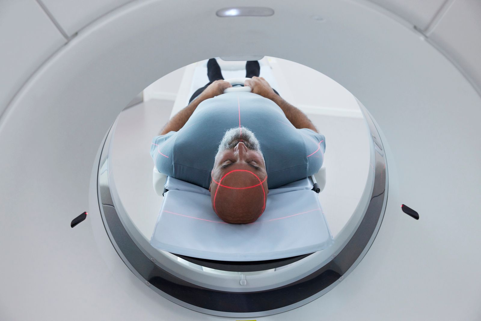
Center of Excellence for Pituitary Surgery
At Hoag’s Skull Base and Pituitary Tumor Program, patients receive world-class care from a team of highly specialized experts who redefine what’s possible in neurosurgery. Our fellowship-trained neurosurgeons, otolaryngologists, neurologists, neuro-ophthalmologists, neuroradiologists, and endocrinologists collaborate to deliver the safest, most effective treatment for even the most complex tumors. With decades of collective experience, our team has helped advance minimally invasive surgical techniques, prioritizing precision, safety, and patient recovery. Most procedures are performed through tiny incisions—or even through the natural passageways of the body, such as the nostrils—reducing pain, scarring, and recovery time. At Hoag, patients can trust that they are in the hands of pioneers who combine compassion, innovation, and advanced technology to achieve the best possible outcomes and quality of life.

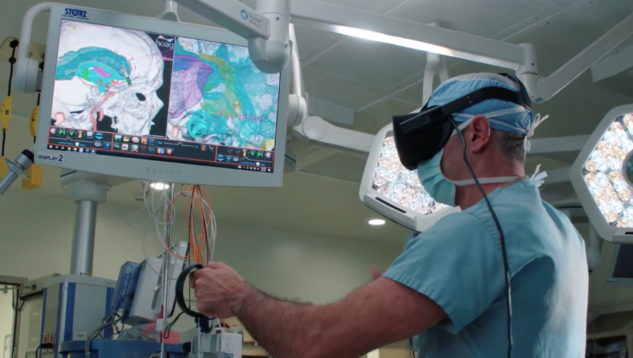
Precision in Motion: How Virtual & Augmented Reality Are Transforming Brain Surgery

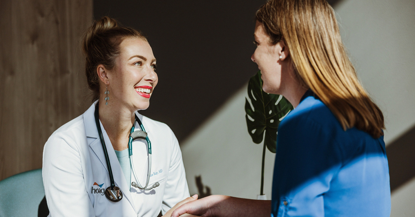
Where Compassion Meets Innovation: A New Era of Patient Comfort
Healing starts with humanity. From our Styled for Surgery program to allowing patients to wear their own clothes, Hoag’s Skull Base and Pituitary Program blends world-class neurosurgery with a hospitality-inspired experience that helps you feel like yourself again. At Hoag, we understand that healing after brain tumor surgery isn’t just physical —it’s emotional, too. That’s why we’re offering a complimentary haircut service for anyone undergoing brain surgery. You’ll meet with Loni Kohlmyer, our licensed stylist, who will create a thoughtful hairstyle to gently conceal your incision, helping you move forward with comfort, confidence, and dignity. The day before surgery, Dr. Goldschmidt will mark the incision site. Then you’ll sit down with Loni to plan a look that feels right for you, showing as much or as little as you choose. Because healing should be personal, and you deserve to feel like you.

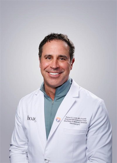
Meet Dr. Louis, MD, FAANS
Dr. Robert Louis, Empower360 Endowed Chair in Skull Base and Minimally Invasive Neurosurgery, shares his innovative approach to patient care. In this video, he highlights the importance of minimally invasive techniques in brain surgery and the critical role of patient education in achieving optimal outcomes. Using advanced VR and AR technology, Dr. Louis guides patients and their families through the surgical journey, mirroring the same pathways used in the operating room with augmented reality overlays on his microscope.
Patient Testimonial Carousel
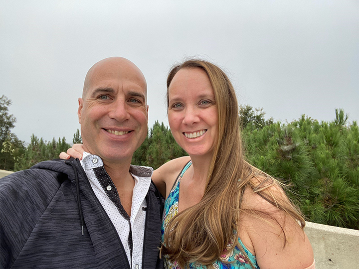
““Hoag is bar-none the best hospital anywhere [...]. On my last day there, she wished me well. Everyone there made me feel like a person, someone who needed help and was seen. I’ve had other surgeries in my life, and that’s not normally how you feel.” ”

Your care starts here
1
Find the right provider
Get care from medical providers that fit your needs in a location near you.
Find a provider2
3
Get in touch
Didn’t see what you’re looking for? Reach out and we’ll make sure you get what you need.
Contact usType of Conditions
Common types of tumors addressed in skull base surgery include meningiomas, which arise from the meninges covering the brain. These tumors can vary in their growth patterns and potential impact on surrounding structures, making surgical intervention complex and delicate.
The majority of pituitary tumors are considered benign (or non-cancerous) because they do not spread to other parts of the body. However, benign tumors can still cause significant health issues due to their location near the brain and optic pathway.
Characteristics of pituitary tumors include:
Usually slow growing
Most often noncancerous
Can grow quite large
Can present with headaches, visual loss and hormonal imbalances
Other pituitary tumors can cause excessive or lessened production of the hormones that regulate necessary body functions. Treatment of such pituitary tumors can include removal of the tumor, control of tumor growth, or tumor management with medication.
Types of Skull Base Tumors
These rare tumors arise from remnants of the notochord and are typically located at the base of the skull or along the spine. They tend to grow slowly but can be aggressive and invasive, often requiring complex surgical approaches.
These malignant tumors arise from cartilaginous tissue and are less common. They often require aggressive treatment due to their potential to invade surrounding structures.
A colloid cyst is a slow-growing, benign cyst that forms near the center of the brain, often at the third ventricle. It can block the normal flow of cerebrospinal fluid, leading to headaches, dizziness, or in rare cases, sudden neurological symptoms. Minimally invasive surgery is often used to remove the cyst and restore fluid balance.
An epidermoid cyst of the brain is a rare, benign tumor that develops from skin-like cells trapped during brain formation. These cysts can grow slowly and press on nerves or brain tissue, causing symptoms like facial pain, hearing loss, or imbalance. Surgical removal is typically recommended for relief and to prevent recurrence.
Gliomas are a group of primary brain tumors that develop from glial cells — the supportive cells that surround and protect neurons in the brain. These tumors can vary in aggressiveness, growth rate, and treatment approach, depending on their type and grade. Treatment often includes a combination of surgery, radiation therapy, and chemotherapy, guided by advanced imaging and molecular diagnostics to tailor care for each patient.
Glioblastoma: The most aggressive form of glioma, glioblastoma tends to grow and spread rapidly. Treatment focuses on maximal surgical removal followed by radiation and chemotherapy to slow progression and improve quality of life.
Ependymoma: Arising from the cells lining the fluid-filled spaces of the brain and spinal cord, ependymomas can block cerebrospinal fluid flow and increase pressure in the brain. Treatment typically involves surgery and radiation therapy.
Astrocytoma: These tumors form from star-shaped glial cells called astrocytes and range from slow-growing (low-grade) to aggressive (high-grade) types. Treatment depends on tumor location and grade, balancing tumor control with preservation of neurological function.
Oligodendroglioma: Originating from cells that produce myelin, these tumors are generally slower-growing and often more responsive to treatment. Genetic testing helps guide therapy, as certain mutations can predict better treatment outcomes.
Hemangioblastomas are rare, benign vascular tumors that occur in the brain or spinal cord. They may cause headaches, problems with balance, or other neurological symptoms depending on their location. Surgical removal is often curative, and ongoing monitoring is important, especially for patients with genetic conditions such as von Hippel-Lindau disease.
These are the most common primary tumors of the skull base. They are usually benign and originate from the meninges. Symptoms can vary depending on their location and size but may include headaches, seizures, or neurological deficits.
Metastatic brain tumors occur when cancer from another part of the body spreads to the brain. Symptoms depend on tumor size and location and can include headaches, seizures, or neurological changes. Treatment options include surgery, radiation, targeted therapy, or a combination to control growth and relieve symptoms.
Olfactory neuroblastoma, also known as esthesioneuroblastoma, is a rare cancer that develops in the upper nasal cavity near the smell nerves. Symptoms can include nasal congestion, nosebleeds, or loss of smell. Treatment often combines surgery, radiation therapy, and sometimes chemotherapy.
These are rare tumors that originate from paraganglia, collections of nerve cells. They are usually benign but can occasionally be malignant.
Sinonasal carcinoma refers to a group of cancers that develop in the nasal cavity or sinuses. These tumors may cause nasal congestion, facial pain, or swelling. Early diagnosis is critical, as treatment typically involves surgery followed by radiation or chemotherapy to preserve function and control disease.
Subependymomas are rare, typically slow-growing tumors that develop from the ependymal cells lining the ventricles of the brain or the central canal of the spinal cord; they are usually noncancerous. When symptoms occur, they may include headaches, balance issues, or changes in vision due to blockage of cerebrospinal fluid flow. Treatment often depends on the size and location of the tumor — some may only require observation, while others are surgically removed to relieve pressure and prevent complications.
These benign tumors develop on the vestibular nerve, which connects the inner ear with the brain. They can cause hearing loss, tinnitus, and balance issues as they grow.
Types of Pituitary Tumors
These are generally benign tumors that occur in the pituitary gland. They can affect hormone levels, leading to various systemic symptoms like vision problems, headaches, or hormonal imbalances
Acromegaly is a rare disorder caused by the overproduction of growth hormone (GH). This excess GH leads to abnormal growth of bones, cartilage, and soft tissues in adults.
Cushing's disease is a specific type of Cushing's syndrome, a condition caused by prolonged exposure to high levels of the hormone cortisol. In Cushing's disease, the culprit is a non-cancerous tumor in the pituitary gland that produces too much adrenocorticotropic hormone (ACTH). This excess ACTH then signals the adrenal glands to produce excessive cortisol.
These benign tumors develop near the pituitary gland and can affect its function, leading to hormonal imbalances and vision problems.
A Rathke cleft cyst is a benign, fluid-filled cyst that develops near the pituitary gland. Though noncancerous, it can cause headaches, vision changes, or hormonal imbalances when it grows large enough to press on nearby structures. Treatment may involve monitoring or surgical drainage to relieve symptoms.
Types of Vascular Compression Syndromes
Characterized by sudden, severe facial pain, trigeminal neuralgia occurs when a blood vessel compresses the trigeminal nerve, which carries sensation from the face to the brain. Treatment may include medication, nerve blocks, or surgical decompression to relieve pressure on the nerve.
This condition causes involuntary twitching or contraction of muscles on one side of the face due to irritation of the facial nerve. Microsurgical decompression or targeted Botox® injections can help restore normal facial movement and reduce discomfort.
Involving sharp pain in the throat, tongue, or ear, this rare condition results from compression of the glossopharyngeal nerve. Treatment options include medications to control pain or surgical decompression to relieve nerve pressure.
This structural defect occurs when brain tissue extends into the spinal canal, often disrupting cerebrospinal fluid flow and causing headaches, neck pain, or balance issues. Surgical treatment can restore normal fluid movement and relieve symptoms.
SSCD occurs when a small hole develops in the bone overlying the inner ear canal, leading to hearing loss, dizziness, or sound sensitivity. Surgical repair can correct the defect and significantly improve balance and hearing.
Skull Base Surgery
Skull base neurosurgery is a specialized surgical approach focused on the area at the bottom of the skull, which includes complex anatomical structures like nerves, blood vessels, and the brain. This type of surgery is often employed to remove tumors, repair abnormalities, or address conditions affecting the base of the skull and surrounding areas. It requires a high level of expertise due to the intricate nature of the region and the proximity to critical brain structures.
Techniques used in these surgeries can be either open or minimally invasive. Open surgeries involve making an incision to access the skull base, while minimally invasive techniques use endoscopes through the nose or small openings. The choice of technique depends on the location and type of the condition. The surgery aims to remove or repair the problematic area while minimizing damage to nearby critical structures like the brain, cranial nerves, and blood vessels. Advanced imaging and navigation technologies often assist surgeons in achieving precise results.
Virtual & Augmented Reality in Neurosurgery
At Hoag, advanced technology meets surgical artistry. Using the Precision Virtual Reality® platform by Surgical Theater, our neurosurgeons transform traditional MRI and CT scans into an immersive 3D model of your brain. This allows our experts to study every angle of your anatomy and rehearse the procedure virtually before entering the operating room.
Patients and their families are invited to “walk the surgery” alongside their surgeon, gaining a clear understanding of where the tumor is located and how it will be safely removed. This approach not only enhances precision but also eases anxiety, turning fear of the unknown into confidence and collaboration.
During surgery, the same technology transitions into an augmented reality (AR) experience, overlaying the surgical path within the surgeon’s microscope. It’s like having X-ray vision, offering unparalleled accuracy and minimizing risk to surrounding tissue.
Hoag remains one of the nation’s highest-volume centers using AR in neurosurgery and continues to advance the field under the leadership of Dr. Louis, Empower360 Endowed Chair in Skull Base and Minimally Invasive Neurosurgery.
Inside the Surgical Theater
The Skull Base and Pituitary Tumor team uses the patient's MRI images to create a virtual map of the brain. Using this technique allows the neurosurgeon to see exactly where the tumor lies and the surrounding structures that may be impacted during surgery. Once the full view of the skull and its structures has been made into the model, the surgeon can map out the surgical path that will allow for the least complications and best outcomes. This is also when the patient and their families can view the surgical path and discuss with their surgeon any questions they may have.

Pre-surgery Rehearsal
After the surgical path has been confirmed, the neurosurgeon can then "practice" the surgery in a virtual format. This allows the surgeon to know exactly where the structures lie, where they can enter the brain and what obstructions they may encounter. Practicing in a virtual environment allows the surgeon to become comfortable with the unique structure and allows for consistency in approach.

Inside the OR
After mapping the brain, reviewing the surgical pathways and rehearsing the surgery, the neurosurgeon is ready to begin removal of the obstruction. The same image that was used in the virtual reality setting is now overlaid in the microscope. This augmented reality allows the surgeon to have "X-ray vision" to see the structures before and during entry. This method allows for precision and best outcomes for the patient.

Skull Base Surgery Program
Surgical treatment of a pituitary tumor is often necessary when it causes any variety of hormone problems, compressing important nerves and arteries at the base of the brain, or when the tumor is putting pressure on optic nerves, bringing on loss of vision. Surgical success rates depend on the location, type and size of tumor, and whether it has spread to surrounding areas.
There are two types of pituitary surgery including the minimally invasive endoscopic transsphenoidal approach and the transcranial approach, also known as craniotomy.
Revolutionizing Preparation for Brain Surgery
Hoag became Orange County’s first and only hospital to treat neurosurgery patients using the same sophisticated software platform that powers today’s highly immersive and realistic computer games.
Developed by a company called Surgical Theater, Surgical Navigation Advanced Platform, or SNAP software blends medical imaging with the latest gaming technology and 3D virtual reality systems. The result is an extraordinarily advanced imaging tool that revolutionizes the way neurosurgeons prepare for and perform delicate cranial procedures.
Types of Surgery
Minimally invasive endoscopic transsphenoidal surgery enables the doctor to remove the tumor through the nose and sinuses, without the need for an external incision. The technique uses the nostrils as a natural portal to the pituitary gland without the need for an external incision. The doctor works through the nostrils with a tiny camera and a light called an endoscope. An opening is made in the nasal septum, sphenoid sinus, and sella to reach the pituitary. Once the pituitary is exposed, the doctor removes the tumor. Remaining areas of the brain are unaffected, and the procedure leaves no visible scar.
This procedure is conducted with a team of neurosurgeons and ear, nose, and throat (ENT) surgeons.
The vast majority of pituitary tumors can safely be removed through the endoscopic, transsphenoidal route. Exceedingly large tumors may be difficult to remove endoscopically, however, especially in cases where the tumor has spread to adjacent nerves or brain tissue. In these rare cases, a more extensive operation may be required that uses a craniotomy combined with skull base approaches.
Supraorbital eyebrow craniotomy is a safe and effective approach used to remove metastatic brain tumors, gliomas, meningiomas and craniopharyngiomas located along the subfrontal section of the brain or around the pituitary gland, which are close to the optic nerve or vital arteries. This approach requires a small incision within the eyebrow line.
For prolactinoma, surgery may not be required as this type of tumor responds well to medication and close observation with periodic MRI to monitor tumor growth.
Where Hospitality Meets Healthcare
At Hoag, we believe that exceptional care extends beyond the operating room. Our vision is to combine hospitality and healthcare, ensuring that every patient feels seen, supported, and respected throughout their journey.
Through our flagship Styled for Surgery program, patients receive personalized haircuts before surgery designed to discreetly hide incision lines, helping you return to life feeling confident and whole. You’ll also have the comfort of wearing your own clothes, skipping unnecessary pre-surgery testing the day of, and avoiding the ICU after most craniotomies.
These thoughtful touches aren’t just about comfort; they’re about dignity. By minimizing the visible and emotional impact of surgery, we help you focus on what matters most: healing and getting back to yourself.
Because at Hoag, your care doesn’t end when surgery does;it begins with how we make you feel every step of the way.
Meet Hoag's Skull Base & Pituitary Tumors Team
Skull Base & Pituitary Tumors Surgeons

Robert G. Louis Jr, MD
Director of Brain Tumor Program
Timothy H. Lucas Jr, MD
Chief of Neurosurgery

Ezequiel D. Goldschmidt, MD
Brain and Skull Base Tumor Neurosurgery

Timothy F. Kelley, MD
Program Director, Hoag Head & Neck Cancer
Neuro-Oncologist

Simon Khagi, MD
Medical Director, Neuro-Oncology
Radiation Oncologists

Brian S. Kim, MD
Radiation Oncology

Shane A. Lloyd, MD
Radiation Oncology

Jennifer Lozano, BSN, RN
Clinical Nurse Navigator
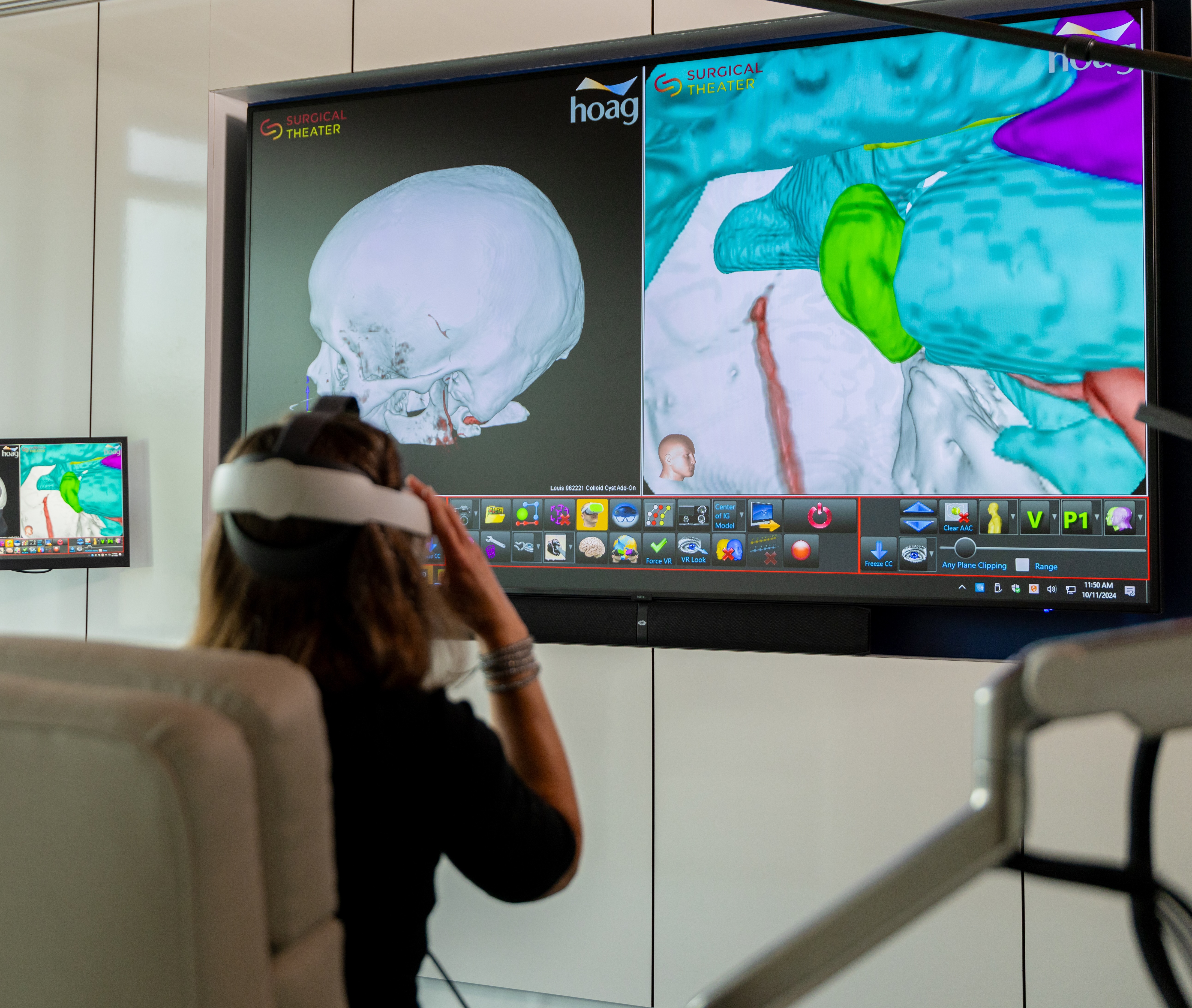
Stay up-to-date on the latest news from Hoag
By submitting this request, you agree to receive communications from Hoag and accept our Privacy Policy and Terms of Use.


